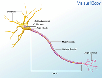
All nervous tissue, from the brain to the spinal cord to the furthest nerve branch, includes cells called neurons. Neurons are charged cells: they conduct electrical signals to pass information through the body. See it in 3D!
System: Nervous
Region: All
Function: They transmit sensory signals and motor commands.
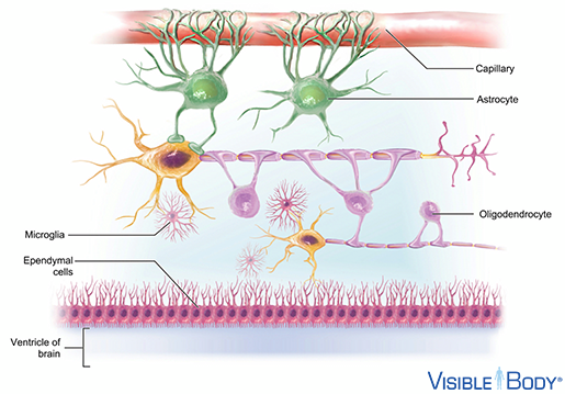
Neuroglia support the neurons and other structures that supply and surround nervous tissue.
System: Nervous
Region: All
Function: Astrocytes, the most common neuroglia in the brain, surround capillaries, maintain a barrier between the bloodstream and the neurons, and actively control what gets through that barrier. Other neuroglia, including microglia, ependymal cells, and oligodendrocytes, maintain neuronal homeostasis, remove pathogens, circulate cerebrospinal fluid, protect neurons, and affect their signaling speed.
Neurotransmitters are chemicals released at synapses that regulate the activity of muscles, glands, and other neurons.
System: Nervous
Region: All
Function: Excitatory neurotransmitters encourage the transmission of an action potential, while inhibitory neurotransmitters inhibit transmission.
Neurotransmitters travel across synapses, spaces between neurons or between neurons and other body tissues and cells.
System: Nervous
Region: All
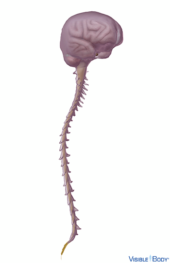
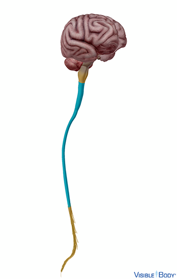
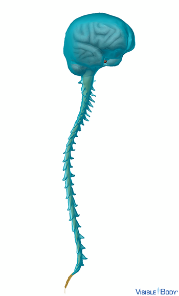
The brain and the spinal cord make up the central nervous system. See it in 3D!
System: Nervous
Region: Head, Neck, Back
Function: The brain and spinal cord (the CNS) function as the control center. They receive data and feedback from the sensory organs and from nerves throughout the body, process the information, and send commands back out.
Medulla spinalis
The spinal cord is an elongated cylinder of neuron cell bodies, bundles of axons and other cells, protected by connective tissue and bone. It connects to the brain at the medulla oblongata and runs down the vertebral column, the hollow tunnel enclosed within the vertebrae of the spine.
System: Nervous
Region: Neck, Back
Function: The spinal cord is part of the central nervous system and serves as a kind of superhighway. Sensory information and motor commands travel up and down, heading to and from the brain.
Pathologies: Arteriovenous malformations, meningitis, multiple sclerosis, neural tube defects, polio and post-polio syndrome, spina bifida, spinal muscular atrophy, syringomyelia
The meninges are the three layers of connective tissue that surround and protect the brain and spinal cord. These are the dura mater, arachnoid mater, and pia mater.
System: Nervous
Region: Head, Neck, Back
Encephalon
The brain—the upper, expanded part of the central nervous system contained within the cranium (neurocranium or braincase)—is a pinkish, soft, ovoid organ that receives sensory input and integrates information to form perception and thought, control activities such as speech and movement, and maintain homeostasis. See it in 3D!
System: Nervous
Region: Head
Pathologies: Alzheimer’s Disease, amyotrophic lateral sclerosis, aphasia, arteriovenous malformation, brain aneurysm, brain cancer, cerebral palsy, Chiari malformation, coma, concussion, Creutzfeldt-Jakob Disease, delirium, dementia, encephalitis, epilepsy, fainting, Huntington’s Disease, hydrocephalus, Lewy body disease, migraine, mild cognitive impairment, neuroblastoma, Parkinson’s Disease, progressive supranuclear palsy, seizures, stroke, Tourette Syndrome, transient ischemic attack, traumatic brain injury
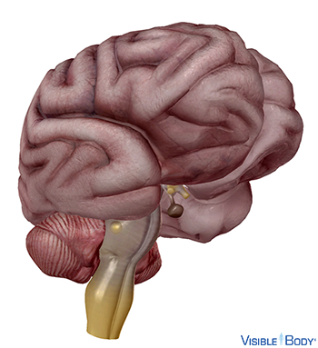
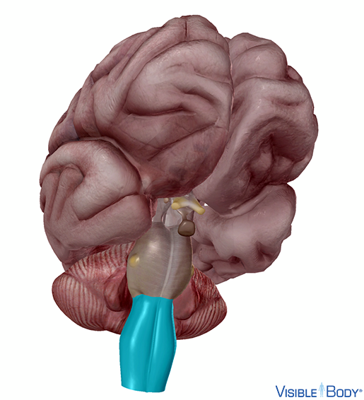
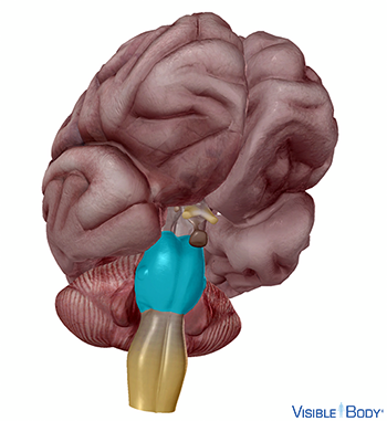
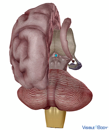
The brainstem connects the spinal cord to the higher-thinking centers of the brain. It consists of three structures: the medulla oblongata, the pons, and the midbrain. See it in 3D!
System: Nervous
Region: Head
Function: Besides relaying sensory and motor signals, the structures of the brain stem direct involuntary functions.
Medulla oblongata
The medulla oblongata is continuous with the spinal cord and connects to the pons above. Both the medulla and the pons are considered part of the hindbrain.
System: Nervous
Region: Head
Function: The medulla handles respiration, digestion, and circulation, and reflexes such as swallowing, coughing, and sneezing.
Pons
The pons (r, l), part of the metencephalon of the hindbrain, bridges the two main function areas of the central nervous system, the “higher” brain centers and the spinal cord.
System: Nervous
Region: Head
Function: The pons helps control breathing rhythms.
Mesencephalon
The midbrain, or mesencephalon, connects the pons to the diencephalon and forebrain.
System: Nervous
Region: Head
Function: The midbrain contributes to motor control, vision, and hearing, as well as vision- and hearing-related reflexes.
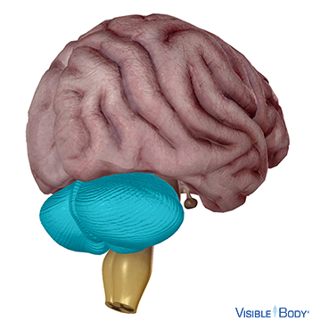
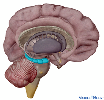
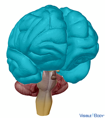
Cerebellum
The cerebellum is the second largest part of the brain. It sits below the posterior (occipital) lobes of the cerebrum and behind the brain stem, as part of the hindbrain. Like the cerebrum, the cerebellum has left and right hemispheres. A middle region, the vermis, connects them. See it in 3D!
System: Nervous
Region: Head
Function: The primary function of the cerebellum is to maintain posture and balance.
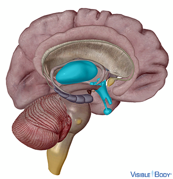
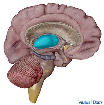
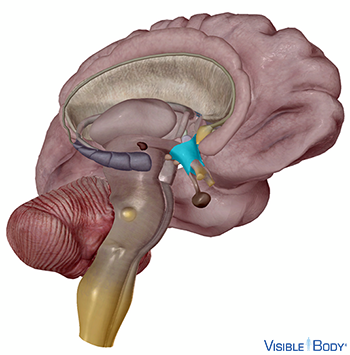
The diencephalon (r, l), a region of the forebrain, comprises the thalamus, the hypothalamus, and the epithalamus.
System: Nervous
Region: Head
Function: The diencephalon mediates sensations, manages emotions, and commands whole internal systems.
Thalamus
The thalamus forms most of the diencephalon. It consists of two symmetrical egg-shaped masses, with neurons that radiate out through the cerebral cortex.
System: Nervous
Region: Head
Function: Sensory data floods into the thalamus from the brain stem, along with emotional, visceral, and other information from different areas of the brain. The thalamus relays these messages to the appropriate areas of the cerebral cortex.
Hypothalamus
The hypothalamus helps to process sensory impulses of smell, taste, and vision. It manages emotions such as pain and pleasure, aggression and amusement. The hypothalamus is also our visceral control center, regulating the endocrine system and internal functions that sustain the body day to day. See it in 3D!
System: Nervous, Endocrine
Region: Head
Function: It translates nervous system signals into activating or inhibiting hormones that it sends to the pituitary gland. It signals sleep cycles and other circadian rhythms, regulates food consumption, and monitors and adjusts body chemistry and temperature.
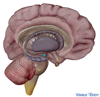
Epiphysis cerebri
The pineal gland (r, l), also called the pineal body, epiphysis cerebri, or epiphysis, is a small reddish-gray body in the epithalamus of the diencephalon (r, l). One region of the pineal gland, the suprachiasmatic nucleus, is often referred to as the “biological clock.” See it in 3D!
System: Nervous, Endocrine
Region: Head
Function: The melatonin secreted by the pineal gland contributes to the regulation of the diurnal cycle.
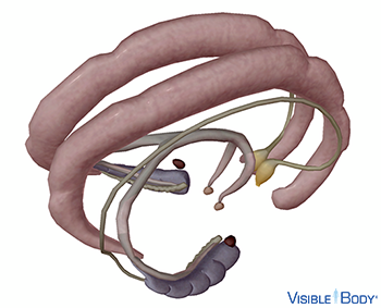
The limbic system forms two paired rings within the brain, consisting of the hippocampus, the amygdala, the cingulate gyrus, and the dentate gyrus, along with other structures and tracts. As with other brain segments, the limbic system is involved in multiple nervous system functions and levels of activity. See it in 3D!
System: Nervous
Region: Head
Function: It helps to process both memory and olfaction—our sense of smell—and it manages a range of emotions.
Hippocampus
The C-shaped hippocampus (r, l) is a structure of the limbic system found in the medial temporal lobe. See it in 3D!
System: Nervous
Region: Head
Function: It plays a role in consolidation of long-term memory.
Amygdala
The amygdala (r, l), a structure of the limbic system, is an ovoid gray mass located on the anterior surface of the hippocampus.
System: Nervous
Region: Head
Function: It is involved in memory and emotion and the linking of the former to the latter.
Cerebrum
The cerebrum is the largest brain structure and part of the forebrain (or prosencephalon). Its prominent outer portion, the cerebral cortex, not only processes sensory and motor information but enables consciousness, our ability to consider ourselves and the outside world. The cortex tissue consists mainly of neuron cell bodies, and its folds and fissures (known as gyri and sulci) give the cerebrum its trademark rumpled surface. The cerebral cortex has a left and a right hemisphere. Each hemisphere can be divided into four lobes: the frontal lobe, temporal lobe, occipital lobe, and parietal lobe.
System: Nervous
Region: Head
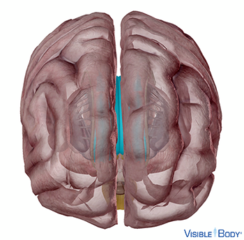
Corpus callosum
The corpus callosum (r, l), a central white commissure that crosses the midline of the brain, connects corresponding gray matter regions of two hemispheres of the cerebrum, allowing communication between the two sides of the brain and enabling integrated function.
System: Nervous
Region: Head
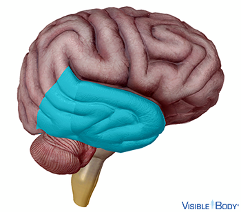
Lobus frontalis
The frontal lobe (r, l), the most anterior of the cerebrum’s four lobes, extends from the anteriormost region of the cerebrum to the central sulcus.
System: Nervous
Region: Head
Function: The speech center, known as Broca’s area, is in the frontal lobe. Other regions of the frontal lobe are involved in what are considered to be “higher functions” such as planning and long-term memory.
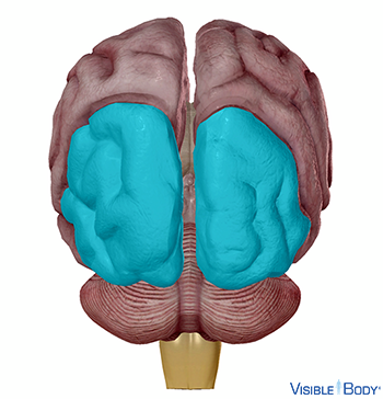
Lobus temporalis
The temporal lobe (r, l) of the cerebrum contains an auditory cortex that receives input from the cochlear nerve, and association areas that integrate auditory, olfactory, and complex pattern perception.
System: Nervous
Region: Head
Lobus parietalis
The parietal lobe (r, l) of the cerebrum integrates sensory information and plays a role in spatial perception.
System: Nervous
Region: Head
Function: The postcentral gyrus (r, l), or primary somatosensory cortex, is located in the parietal lobe. It receives sensory information and gives the perception of touch. Other areas of the parietal lobe process awareness of body’s motion, touch, perception of temperature, pain, and movement.
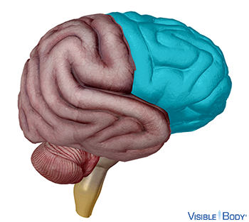
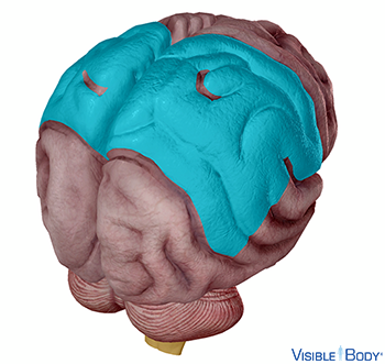
Lobus occipitalis
The occipital lobe is the posterior lobe of the cerebrum that receives input from the eye and processes visual perceptions.
System: Nervous
Region: Head
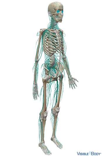
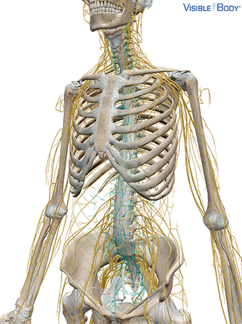
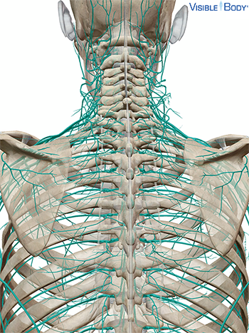
The peripheral nervous system (PNS) consists of all of the nerves and ganglia outside the central nervous system that connect it to tissues throughout body regions. The nerves that branch off the central nervous system are known as the cerebrospinal nerves. There are 43 on each side: 12 cranial and 31 spinal.
System: Nervous
Region: All
Function: Each nerve is responsible for relaying sensory information, sending motor commands, or both.
Pathologies: Complex regional pain syndrome
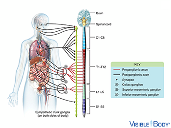
The autonomic nervous system is the division of the peripheral nervous system that regulates involuntary visceral functions such as heartbeat and smooth muscle contraction. The autonomic nervous system is divided into sympathetic and parasympathetic divisions.
System: Nervous
Region: All
Pathologies: Complex regional pain syndrome
Sympathetic nerves originate in the thoracic and lumbar portion of the spinal cord and form a series of ganglia known as the sympathetic chain, as well as three major plexuses: cardiac, celiac, and hypogastric.
System: Nervous
Region: Thorax, Abdomen
Function: Nerve impulses from sympathetic nerves increase heart rate and other body functions in response to an emergency.
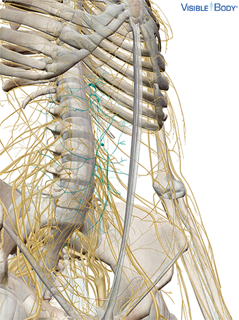
The celiac plexus is the largest of the three sympathetic plexuses of the autonomic nervous system. The celiac plexus is composed of two large ganglia, the celiac ganglia, and a dense network of nerve fibers connecting them together.
System: Nervous
Region: Thorax, Abdomen
Pathologies: Complex regional pain syndrome
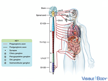
Parasympathetic fibers extend from cranial and sacral nerves, and are responsible for rest functions such as digestion.
System: Nervous
Region: Head, Neck, Thorax, Abdomen
N. spinalis
The 31 pairs of spinal nerves connect tissues in the thorax, abdomen, and limbs to the spinal cord; these nerves contain both sensory and motor fibers and are therefore referred to as mixed.
System: Nervous
Region: Neck, Thorax, Abdomen, Pelvis
Pathologies: Complex regional pain syndrome
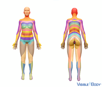
A dermatome is a region of skin that is innervated by the sensory fibers from a single spinal nerve. All the sacral, lumbar, thoracic, and cervical spinal nerves innervate dermatomes except the first cervical spinal nerve (C1), which does not contain any sensory axons.
System: Nervous, Integumentary
Region: All
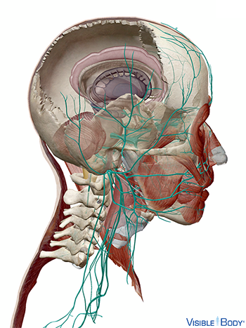
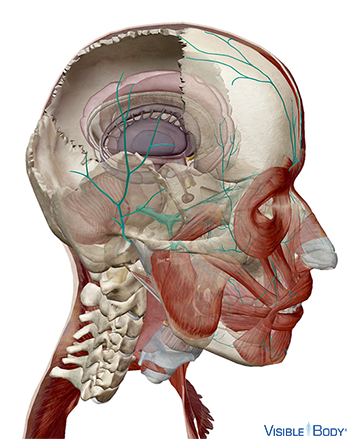
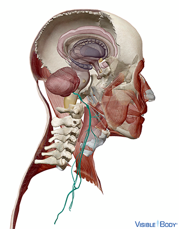
Nn. craniales
Twelve pairs of cranial nerves connect the brain to eyes, ears, and other sensory organs and to head and neck muscles. See it in 3D!
System: Nervous
Region: Head, Neck, Thorax, Abdomen
Pathologies: Complex regional pain syndrome
N. trigeminus
The trigeminal nerves (V) are the largest cranial nerves are the largest cranial nerves. They consist of ophthalmic, maxillary, and mandibular branches that pass sensory and motor signals between the pons and structures of the face.
System: Nervous
Region: Head
Pathologies: Complex regional pain syndrome, trigeminal neuralgia
N. vagus
The vagus nerves transmit sensory and motor impulses between the medulla oblongata and the visceral organs. They pass through the jugular foramina on their way to the thorax and abdomen.
System: Nervous
Region: Head, Neck, Thorax, Abdomen
Pathologies: Complex regional pain syndrome
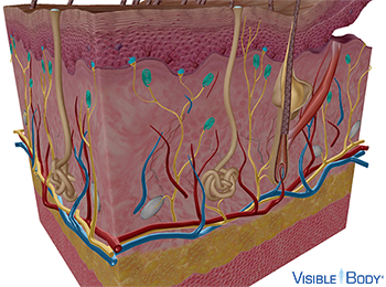
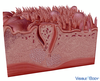
Specialized receptor cells within these layers detect tactile sensations and relay signals through peripheral nerves toward the brain. The presence and location of the different types of receptors make certain body parts more sensitive. Merkel cells, for example, are found in the lower epidermis of lips, hands, and external genitalia. Meissner corpuscles are found in the upper dermis of hairless skin — fingertips, nipples, the soles of the feet.
System: Nervous (Special Senses), Integumentary
Region: All
Pathologies: Chickenpox, eczema, hives, melanoma, psoriasis, skin pigmentation disorders
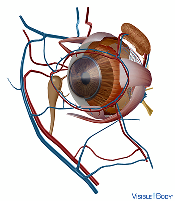
Oculus
The eyes (r, l), spherical sensory organs of the nervous system, are responsible for sight. See it in 3D!
System: Nervous (Special Senses)
Region: Head
Function: The retina is a delicate membrane of nervous tissue containing photoreceptor cells. These cells, the rods and cones, translate light into nervous signals. The optic nerve carries the signals from the eye to the brain, which interprets them to form visual images.
Pathologies: Amblyopia, cataract, color blindness, complex regional pain syndrome, macular degeneration, pinkeye, refractive errors, retinal detachment, rheumatoid arthritis
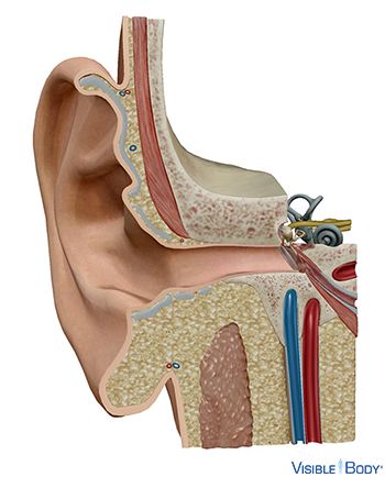
Auris
The ear consists of three sections: the outer (or external ear), the middle ear, and the inner (or internal) ear. See it in 3D!
System: Nervous (Special Senses)
Region: Head
Function: To facilitate hearing, the outer ear collects sound waves and conducts them to the middle ear in the form of vibrations. The auditory ossicles transfer these vibrations to the inner ear. As waves of pressure reach the cochlea, nerve impulses are generated that travel to the brain through the vestibulocochlear nerve (VIII).
Pathologies: Acoustic neuroma, ear infections, Meniere’s Disease, tinnitus
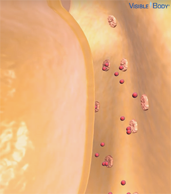
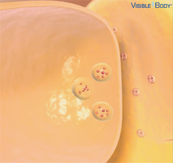
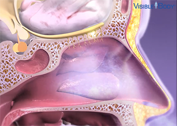
The sense of smell is called olfaction. It starts with specialized nerve receptors located on hairlike cilia in the epithelium at the top of the nasal cavity. When we sniff or inhale through the nose, some chemicals in the air bind to these receptors. That triggers a signal that travels up a nerve fiber, through the epithelium and the skull bone above, to the olfactory bulbs. The olfactory bulbs contain neuron cell bodies that transmit information along the cranial nerves, which are extensions of the olfactory bulbs. They send the signal down the olfactory nerves, toward the olfactory area of the cerebral cortex.
System: Nervous (Special Senses)
Region: Head
Pathologies: Complex regional pain syndrome
Lingua
The tongue is the principal organ of taste and also assists in the mastication and deglutition of food. Small projections called papillae, many of which contain taste buds, cover the dorsal and lateral surface of the tongue. See it in 3D!
System: Nervous (Special Senses), Digestive
Region: Head
Function: When we eat, chemicals from food enter the papillae and reach the taste buds. These chemicals (or tastants) stimulate specialized gustatory cells inside the taste buds, activating nervous receptors.