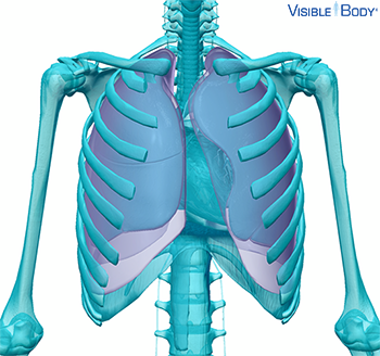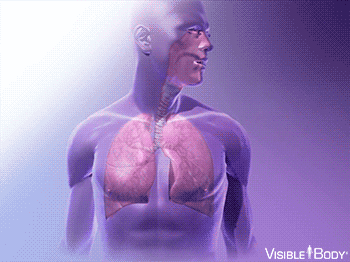
The respiratory system aids in breathing, also called pulmonary ventilation. In pulmonary ventilation, air is inhaled through the nasal and oral cavities (the nose and mouth). It moves through the pharynx, larynx, and trachea into the lungs. Then air is exhaled, flowing back through the same pathway. Changes to the volume and air pressure in the lungs trigger pulmonary ventilation.
System: Respiratory, Muscular
Region: Thorax, Abdomen
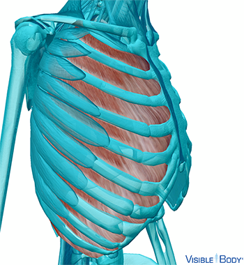
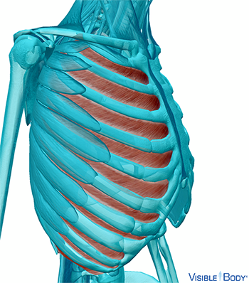
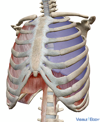
Musculi intercostales externi
The external intercostals (r, l) are muscles of the thorax that extend from the tubercles of the ribs behind to the cartilages of the ribs in front. Their fibers are directed obliquely downward and laterally on the back of the thorax, and downward, forward, and medialward on the front.
System: Muscular
Region: Thorax
Function: Elevates the ribs, aiding in normal inspiration
Pathologies: Muscular dystrophy, myositis
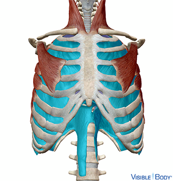
Musculi intercostales interni
The internal intercostals (r, l) are muscles of the thorax that extend from the sternum to the vertebral column. Their fibers are directed obliquely and pass in a direction opposite to those of the external intercostals.
System: Muscular
Region: Thorax
Function: Depresses the ribs, aiding in forced expiration.
Pathologies: Muscular dystrophy, myositis
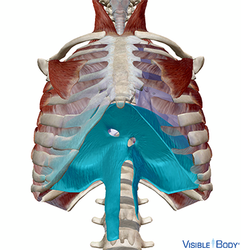
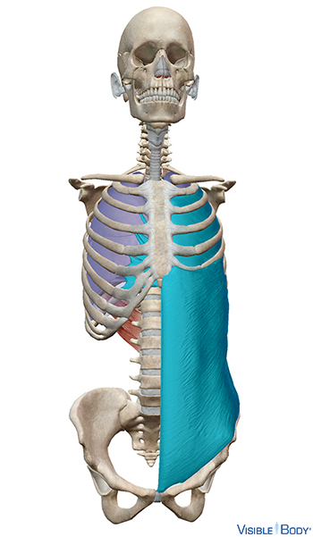
Diaphragma
The diaphragm, a muscle of the thorax, is the principal muscle of inspiration. The diaphragm separates the thoracic and abdominal cavities. See it in 3D!
System: Muscular
Region: Thorax
Function: Expands the thoracic cavity; compresses the abdominal cavity.
Pathologies: Muscular dystrophy, myositis
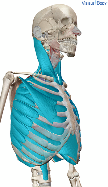
During normal inhalation, the diaphragm and external intercostal muscles contract and the ribcage elevates. As the volume of the lungs increases, air pressure drops and air rushes in.
System: Respiratory, Muscular
Region: Thorax
In forced inhalation, the sternocleidomastoid, scalenes, pectoralis minor, and serratus anterior also contract.
System: Respiratory, Muscular
Region: Neck, Thorax
During normal exhalation, the diaphragm and external intercostals relax. The lungs become smaller, the air pressure rises, and air is expelled.
System: Respiratory, Muscular
Region: Thorax
During forced exhalation, the internal intercostals, transversus thoracis, and abdominal muscles contract.
System: Respiratory, Muscular
Region: Thorax, Abdomen
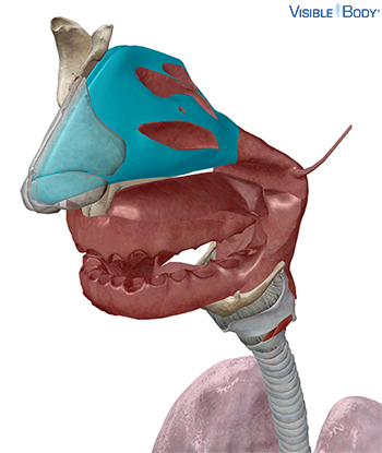
Cavitas nasi
The nasal cavities (r, l) are chambers of the internal nose and function as part of the upper respiratory system. The nasal cavities open in front through the nares, or nostrils; in the back, the nasal cavities connect to the nasopharynx. See it in 3D!
System: Respiratory
Region: Head
Function: Air is inhaled through the nares and warmed as it passes through the nasal cavity.
Pathologies: Allergies, common cold, coronavirus infections
Cartilago septi nasi
The rectangular septal cartilage separates the nasal cavities; it connects to the nasal bones, the greater alar cartilages, and the perpendicular plate of the ethmoid by fibrous connective tissue. See it in 3D!
System: Skeletal
Region: Head
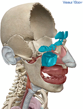
Sinus paranasales
The paranasal sinuses are four paired, air-filled cavities found inside bones of the skull. These sinuses are named for the skull bones that contain them: frontal, ethmoidal, sphenoidal, and maxillary. See it in 3D!
System: Respiratory
Region: Head
Function: Mucosae line the paranasal sinuses and help to warm and humidify the air we inhale.
Pathologies: Allergies, common cold, coronavirus infections, sinusitis
Pharynx
The pharynx is a 12.5-cm conical musculomembranous tube that functions as part of the alimentary canal and as an airway in the upper respiratory system. The pharynx is divided into three segments: the nasopharynx, the oropharynx, and the laryngopharynx. See it in 3D!
System: Respiratory, Digestive
Region: Head, Neck
Function: During respiration, it conducts air between the larynx and trachea (or “windpipe”) and the nasal and the oral cavities.
Pathologies: Allergies, common cold, coronavirus infection, streptococcal infection, whooping cough
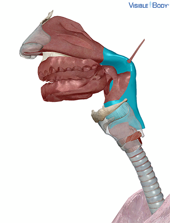
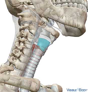
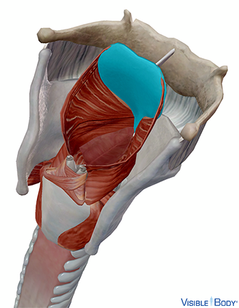
Larynx
The larynx connects the lower part of the pharynx, the laryngopharynx, to the trachea. It keeps the air passages open during breathing and digestion and is the key organ for producing sound. See it in 3D!
System: Respiratory
Region: Neck
Function: Its main function is to provide an airway for breathing.
Pathologies: Allergies, common cold, coronavirus infections, streptococcal infections, whooping cough
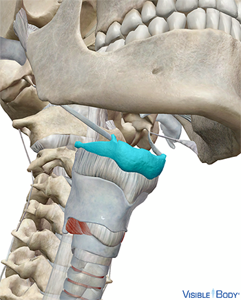
Os hyoideum
In the respiratory system, structures that produce sound depend on the hyoid. The body and the greater horns of the bone serve as attachment points for neck muscles that raise and lower the larynx during speech (as well as during swallowing).
System: Skeletal
Region: Neck
Pathologies: Osteogenesis imperfecta, Paget’s Disease of Bone, rickets
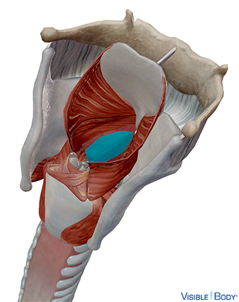
Plicae vocales
The vocal folds, also called the true vocal cords, extend across the cavity of the larynx, the uppermost air passage of the lower respiratory system.
System: Respiratory
Region: Neck
Function: Passing air can vibrate the folds, allowing them to function in the production of sound. The pitch of sounds changes as various muscles regulate the degree of tension of the vocal folds.
Pathologies: Allergies, common cold, coronavirus infections, streptococcal infections, whooping cough
Epiglottis
The epiglottis is one of the nine cartilages that join to form the laryngeal skeleton (also known as the larynx or voice box), which is attached to structures of the axial skeleton. This unpaired structure is leaf-shaped and contains a thin layer of elastic cartilage attached by the thyroepiglottic ligament to the thyroid cartilage. See it in 3D!
System: Skeletal
Region: Neck
Function: Located on the posterior side of the larynx, the epiglottis closes like a trap door as we swallow. This action steers food down the esophagus and away from the windpipe.
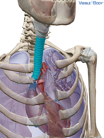
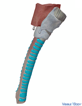
Trachea
The trachea is a tube less than an inch in diameter, covered by cartilaginous rings. It extends from the bottom of the larynx down behind the sternum, until it branches into smaller tubes, the bronchi. See it in 3D!
System: Respiratory
Region: Neck, Thorax
Function: During inhalation, air filtered and warmed by the upper respiratory system passes from the pharynx and larynx into the trachea, then down to the bronchi and into the lungs. Deoxygenated air from the lungs passes back up through the trachea during exhalation.
Pathologies: Allergies, common cold, coronavirus infections, streptococcal infections, whooping cough
Cartilagines tracheales
The tracheal cartilages are stacked horizontally and separated by narrow intervals. The number of cartilages varies from 16 to 20; each forms an incomplete, crescent-shaped ring around the frontal (anterior) two-thirds of the tube.
System: Respiratory
Region: Neck, Thorax
Function: The cartilaginous rings support the tube of the trachea and prevent it from over-expanding or from collapsing, like when you suck on a straw too hard. They are C-shaped, with a gap on the posterior side. This allows the trachea to bend when the esophagus presses against it as food is swallowed.
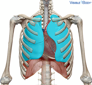
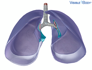
Pulmones
The lungs are responsible for gas exchange between the air we breathe and our bodies. They are protected inside the thoracic cage. The left lung has two lobes and is slightly smaller in volume than the right. It curves in at the cardiac notch to accommodate the heart. The right lung has three lobes. It is slightly shorter, because the diaphragm muscle sits higher below it to accommodate the liver. See it in 3D!
System: Respiratory
Region: Thorax
Pathologies: Acute bronchitis, allergies, asthma, chronic bronchitis, collapsed lung, common cold, COPD, coronavirus infections, cystic fibrosis, emphysema, mesothelioma, pneumonia, pulmonary embolism, pulmonary fibrosis, pulmonary hypertension, respiratory failure, rheumatoid arthritis, tuberculosis, whooping cough
Pleurae
The pleurae are delicate, double-layered serous membranes that cover the lungs.
System: Respiratory
Region: Thorax
Function: The space between the two pleural layers is called the pleural cavity. It contains pleural fluid which provides lubrication to prevent friction as the lungs move and rub against the thoracic wall.
Pathologies: Allergies, common cold, coronavirus infections
Ligamentum pulmonale
The pulmonary ligaments are mesenteric folds on each lung, formed by the union of the visceral and parietal pleurae at their lower borders.
System: Respiratory
Region: Thorax
Function: The pulmonary ligaments help to keep the lower parts of the lungs in position.
Pathologies: Allergies, common cold, coronavirus infections
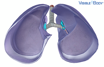
Hilum pulmonis
The hilum (r, l) is a triangular depression where the structures that form the root of the lung enter and leave. Structures entering at the hilum include the bronchus, the pulmonary artery, the pulmonary veins, the bronchial arteries and veins, the pulmonary nerve plexuses, and lymphatic vessels, among others.
System: Respiratory
Region: Thorax
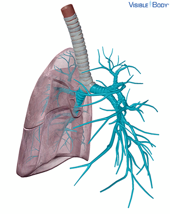
Bronchus
The bronchi and its subdivisions (r, l) are the major airways of the lower respiratory system. The tubes of the primary bronchi branch off from the bottom of the trachea. These branches subdivide further into secondary and tertiary bronchi and then into the bronchioles.
System: Respiratory
Region: Thorax
Function: These progressively smaller airways deliver oxygen-rich air from the trachea to the lungs. During exhalation, deoxygenated air (now rich with carbon dioxide) leaves the lungs by the reverse route.
Pathologies: Acute bronchitis, allergies, asthma, chronic bronchitis, common cold, coronavirus infections
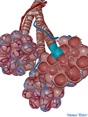
Bronchiolus
As a tertiary bronchus branches within its segment, it gives rise to lobular bronchioles. Lobular bronchioles branch into terminal bronchioles, which mark the end of the conduction zone. Terminal bronchioles branch into respiratory bronchioles, which are the smallest bronchioles and the first portion of the respiratory zone.
System: Respiratory
Region: Thorax
Relaxation of smooth muscle in the bronchioles during exercise causes them to dilate—this bronchodilation allows greater ventilation. Allergic reactions and histamines cause the opposite effect, known as bronchoconstriction.
System: Respiratory
Region: Thorax
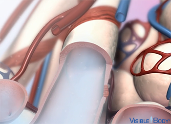
Ductus alveolaris
The respiratory bronchioles directly service alveoli via thin airways called alveolar ducts.
System: Respiratory
Region: Thorax
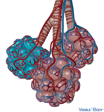
Clusters of alveoli, located at the ends of alveolar ducts, are called alveolar sacs.
System: Respiratory
Region: Thorax
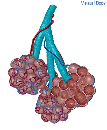
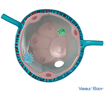
Alveolus
Alveoli are tiny, delicate air sacs in which the main function of the respiratory system occurs: carrying out gas exchange to bring oxygen into the body and remove carbon dioxide. The alveolar wall is the tissue of the alveolus and the space inside the alveolar wall is referred to as the alveolar cavity.
System: Respiratory
Region: Thorax
Type I alveolar cells, also known as squamous alveolar cells or Type I pneumocytes, are simple squamous epithelial cells in the lining of the alveoli of the lungs. They cover 90% of the alveolar surface and are extremely thin.
System: Respiratory
Region: Thorax
Function: Their thinness reduces the diffusion distance for gas exchange, and thus, they enable gas exchange between the alveoli and the blood.
Type II alveolar cells, also known as septal cells or Type II pneumocytes, are epithelial cells in the lining of the alveoli of the lungs. They cover less than 10% of the alveolar surface.
System: Respiratory
Region: Thorax
Function: Their major function is to secrete pulmonary surfactant.
Alveolar macrophages are leukocytes in the alveoli. They are also referred to as dust cells because they engulf very small inhaled particles that reach the alveoli. Alveolar macrophages can be coughed up in the sputum, and their appearance can be diagnostically important.
System: Respiratory
Region: Thorax
Pulmonary surfactant is a fluid composed of phospholipids and proteins that coats the inner surfaces of alveoli.
System: Respiratory
Region: Thorax
Function: It serves to decrease surface tension in the alveoli.
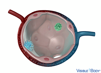
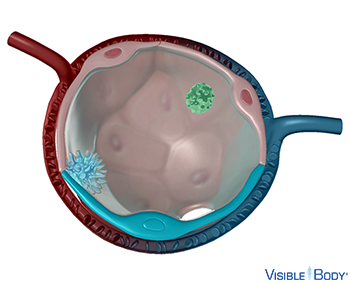
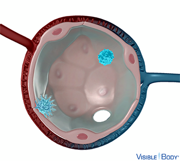
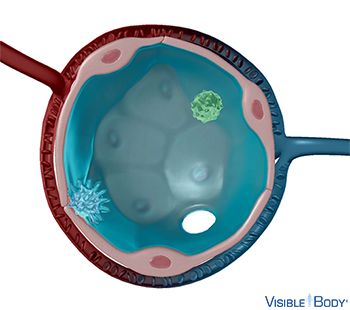
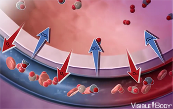
In the lungs, the pulmonary arteries branch into arterioles and then into networks of pulmonary capillaries. Gases are exchanged across the walls of these capillaries, and the oxygen content of the blood rises. The pulmonary capillaries form very thin plexuses immediately beneath the epithelium in the walls and septa of the alveoli and infundibula. The pulmonary capillaries are drained by the pulmonary veins.
System: Circulatory, Respiratory
Region: Thorax
During inhalation, the alveoli fill with air from the bronchioles. Oxygen diffuses through the alveoli into networks of pulmonary capillaries that surround them, and is pumped through the bloodstream. Carbon dioxide from deoxygenated blood diffuses from the capillaries into the alveoli, and is expelled through exhalation.
System: Respiratory
Region: Thorax
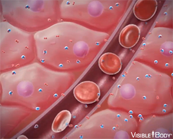
The bloodstream delivers oxygen to cells and removes waste carbon dioxide through internal respiration, another key function of the respiratory system. In this respiratory process, red blood cells carry oxygen absorbed from the lungs around the body, through the vasculature. When oxygenated blood reaches the narrow capillaries, the red blood cells release the oxygen. It diffuses through the capillary walls into body tissues. Meanwhile, carbon dioxide diffuses from the tissues into red blood cells and plasma. The deoxygenated blood carries the carbon dioxide back to the lungs for release.
System: Respiratory
Region: Thorax