Primary endocrine organs secrete hormones that chemically regulate body functions.
System: Endocrine
Region: Head, Neck, Abdomen
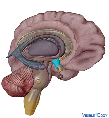
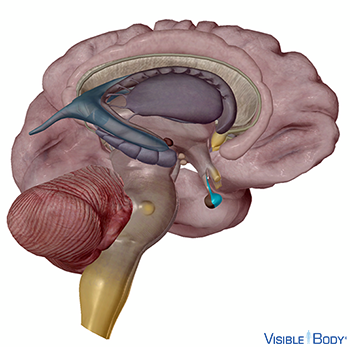
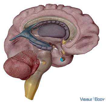
Hypothalamus
The hypothalamus connects the nervous system to the endocrine system. See it in 3D!
System: Endocrine, Nervous
Region: Head (Brain)
Function: It receives and processes signals from other brain regions and pathways and translates them into hormones, the chemical messengers of the endocrine system.
Pathologies: Alzheimer’s Disease, amyotropic lateral sclerosis, aphasia, arteriovenous malformations, brain aneurysm, brain cancer, cerebral palsy, Chiari malformation, coma, concussion, Creutzfeldt-Jakob Disease, delirium, dementia, diabetes insipidus, encephalitis, epilepsy, fainting, Huntington’s Disease, hydrocephalus, Lewy body disease, migraine, mild cognitive impairment, neuroblastoma, Parkinson’s Disease, progressive supranuclear palsy, seizures, stroke, Tourette Syndrome, transient ischemic attack, traumatic brain injury
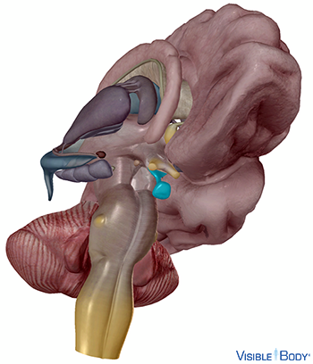
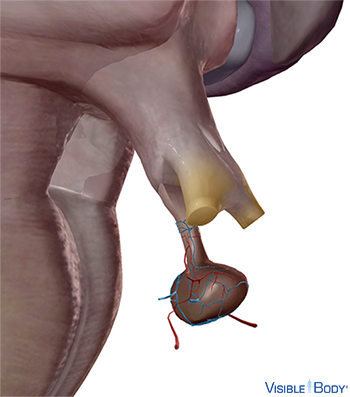
Hypophysis, glandula pituitaria
The pituitary gland (r, l), a small reddish-gray body about 1 cm in diameter, secretes and stores hormones that manage functions of the endocrine system. Attached to the end of the infundibulum of the hypothalamus, the pituitary consists of an anterior lobe and a posterior lobe. See it in 3D!
System: Endocrine
Region: Head (Brain)
Function: The anterior lobe secretes hormones and comprises most of the pituitary’s mass. The posterior pituitary stores and secretes hormones produced by the hypothalamus.
Pathologies: Alzheimer’s Disease, amyotrophic lateral sclerosis, aphasia, arteriovenous malformations, brain aneurysm, brain cancer, cerebral palsy, Chiari malformation, coma, concussion, Creutzfeldt-Jakob Disease, delirium, dementia, diabetes insipidus, encephalitis, epilepsy, fainting, growth disorders, Huntington’s Disease, hydrocephalus, Lewy body disease, migraine, mild cognitive impairment, neuroblastoma, Parkinson’s Disease, progressive supranuclear palsy, seizures, stroke, Tourette Syndrome, transient ischemic attack, traumatic brain injury
Hormones sent from the hypothalamus to the anterior lobe of the pituitary gland function as signals. They stimulate or inhibit the release of anterior pituitary hormones, which regulate endocrine glands and control a range of body functions.
System: Endocrine
Function: Anterior pituitary hormones stimulate growth and control glands throughout the body.
Adrenocorticotropic hormone (ACTH) causes the adrenal glands to produce steroid hormones. These steroid hormones influence the metabolism of glucose, lipids, and proteins, which gives cells energy to resist stress.
System: Endocrine
Source: Anterior pituitary
Target Organ: Adrenal glands
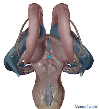
Follicle-stimulating hormone (FSH) targets the gonads in both males and females. In females, it stimulates the ovaries to secrete estrogen and produce oocytes. In males, it stimulates sperm development in the testes.
System: Endocrine
Source: Anterior pituitary
Target Organs: Testes, ovaries
Luteinizing hormone (LH) targets the gonads in both males and females. In males, it stimulates the production of testosterone by the testes. In females, it stimulates ovulation.
System: Endocrine
Source: Anterior pituitary
Target Organs: Testes, ovaries
Human growth hormone (hGH) causes target cells to release insulin-like growth factors (IGFs), which are hormones that promote cell growth and division, glucose release, and protein synthesis.
System: Endocrine
Source: Anterior pituitary
Target Organs: Skeletal muscles, bones, liver
Melanocyte-stimulating hormone (MSH) causes melanocytes in the skin to produce more melanin (pigment).
System: Endocrine
Source: Anterior pituitary (pars intermedialis)
Target Organs: Skin
Prolactin (PRL) induces milk production in mammary glands.
System: Endocrine
Source: Anterior pituitary
Target Organs: Mammary glands
Thyroid-stimulating hormone (TSH) causes the thyroid gland to release hormones that increase metabolism and promote nervous and skeletal system growth.
System: Endocrine
Source: Anterior pituitary
Target Organs: Thyroid gland
The hormones ADH and OXT are produced in the hypothalamus and are stored in the posterior pituitary, from which they are released into circulation.
System: Endocrine
Function: Posterior pituitary hormones regulate water levels and induce labor.
ADH acts on the kidneys, blood vessels, and sweat glands in the skin to reduce water loss throughout the body.
System: Endocrine
Source: Posterior pituitary
Target Organs: Kidneys, blood vessels, sweat glands
OXT factors into pregnancy and nurturing. It causes smooth muscle contractions of the uterus to induce birth. Later it stimulates milk ejection from the mammary glands and promotes bonding between mother and child.
System: Endocrine
Source: Posterior pituitary
Target Organs: Uterus, mammary glands
The system of vasculature that connects the hypothalamus and the anterior pituitary is called the hypophyseal portal system.
System: Circulatory
Region: Head (Brain)
Function: Capillaries from the superior hypophyseal artery surround the hypophysis and collect hypothalamic hormones that are carried to the anterior lobe of the pituitary via the portal system, where they stimulate or inhibit the release of pituitary hormones.
Pathologies: Arteriovenous malformations, diabetes type 1, diabetes type 2
Epiphysis cerebri
The pineal gland is small and pinecone-shaped, located at the posterior of the diencephalon region in the brain. See it in 3D!
System: Endocrine, Nervous
Region: Head (Brain)
Function: The pineal gland secretes melatonin at various levels throughout the day and night; these secretions are thought to contribute to cycles of wake and sleep (circadian cycle).
Pathologies: Alzheimer’s Disease, amyotrophic lateral sclerosis, aphasia, arteriovenous malformations, brain aneurysm, brain cancer, cerebral palsy, Chiari malformation, coma, concussion, Creutzfeldt-Jakob Disease, delirium, dementia, encephalitis, epilepsy, fainting, Huntington’s Disease, hydrocephalus, Lewy body disease, migraine, mild cognitive impairment, neuroblastoma, Parkinson’s Disease, progressive supranuclear palsy, seizures, stroke, Tourette Syndrome, transient ischemic attack, traumatic brain injury
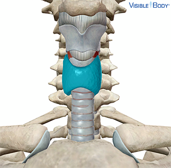
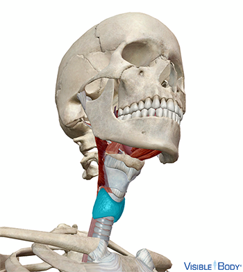
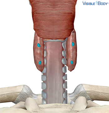
Glandula thyroidea
The thyroid gland sits in the throat region, just below the larynx, served by large arteries with many branches and a dense network of capillaries.
System: Endocrine
Region: Neck
Function: The thyroid gland releases thyroid hormone, in the form of thyroxine (T4) and triiodothyronine (T3), which increase metabolism, glucose use, protein synthesis, and nervous system development. It also releases calcitonin (CT), which promotes calcification of bones. See it in 3D!
Pathologies: Hyperthyroidism, hypothyroidism
Glandula parathyroidea superior, glandula parathyroidea inferior
The parathyroid glands are small, brownish-red structures located between the posterior borders of the lateral lobes of the thyroid gland. Normally, there are four parathyroids: two superior (upper) parathyroid glands (r, l) and two inferior (lower) parathyroid glands (r, l).
System: Endocrine
Region: Neck
Function: They secrete parathyroid hormone (PTH or parathormone), which stimulates bones to release calcium into the blood when blood (calcium) levels are low. PTH also causes the kidneys to reduce calcium secretion into urine to further elevate calcium levels in the blood.
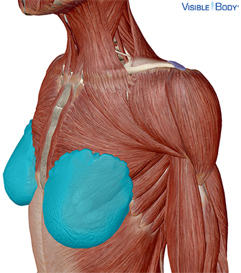
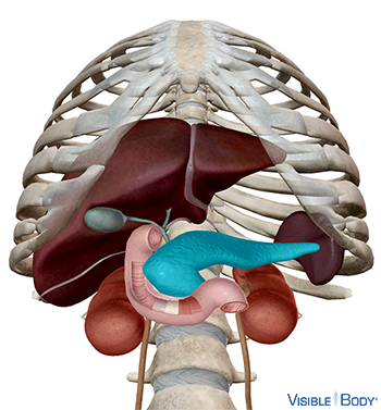
Pancreas
The pancreas is located between the stomach and small intestine. In addition to its digestive functions, it is a gland that secretes hormones necessary to regulate blood glucose levels. See it in 3D!
System: Endocrine, Digestive
Region: Abdomen
Function: When blood sugar is low, alpha cells in the islets release glucagon. Glucagon spurs the liver to break down glycogen and release more glucose into the blood. When blood sugar is high, beta cells in the islets release insulin, which increases glucose reuptake.
Pathologies: Pancreatitis, prediabetes
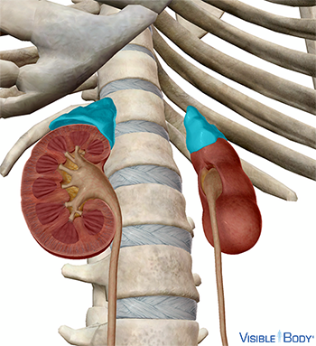
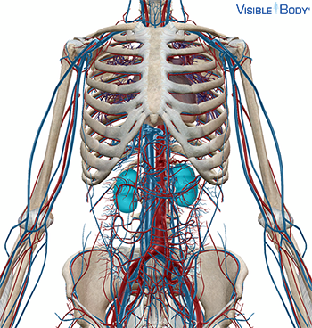
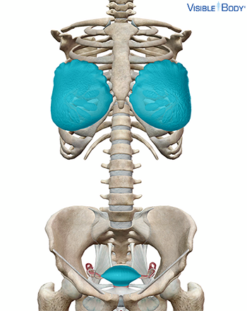
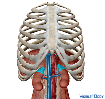
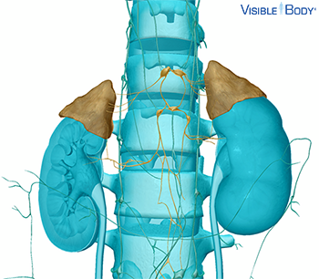
Glandula suprarenalis
The adrenal glands are located superior to the kidneys on either side of the vertebral column. Each adrenal gland consists of an outer cortex and an inner medulla.
System: Endocrine
Region: Abdomen
Function: The adrenal cortex produces three types of steroids. Glucocorticoids, such as cortisol, manage protein and glucose levels. Mineralocorticoids, such as aldosterone, manage the levels of water and salt. Gonadocorticoids are androgens, which can be converted to estradiols. The adrenal medulla produces epinephrine (E) and norepinephrine (NE), which promote the fight-or-flight responses of the sympathetic nervous system during stress.
Pathologies: Pheochromocytoma
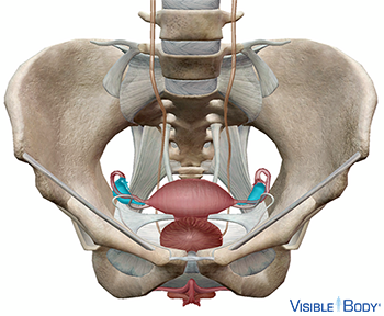
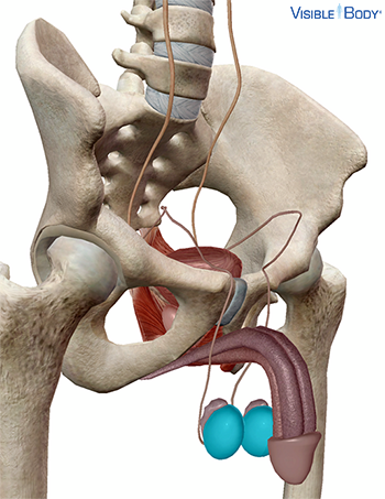
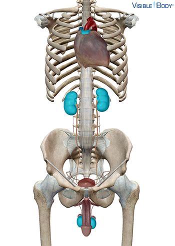
Secondary endocrine organs include the gonads, kidneys, and thymus.
System: Endocrine
Region: Thorax, abdomen
Function: Secondary endocrine organs secrete hormones as a secondary function.
Stress stimulates the adrenal glands to produce hormones that ramp up body activity in the fight-or-flight response. The hypothalamus commands the adrenal glands directly (via nervous signals) to ramp up production of epinephrine and norepinephrine. These hormones promote the “fight-or-flight” response: breathing and heart rate increase and our muscles get a burst of energy. As stress continues into the resistance phase, the pancreas and adrenal glands produce glucagon and steroids that elevate blood glucose to provide more energy.
System: Endocrine