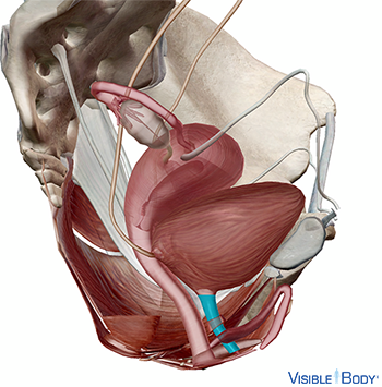
In women, the bladder is located in front of the vagina and below the uterus. The urinary system filters blood to remove waste through urine, which is voided through the urethra. In the female, the urethra passes out of the pelvic cavity through the external urethral orifice.
System: Urinary
Region: Abdomen, Pelvis
Pathologies: Chronic kidney disease, diabetes insipidus, diabetes type 1, diabetes type 2, interstitial cystitis, kidney cysts, kidney failure, kidney stones, overactive bladder, rheumatoid arthritis, urinary incontinence, urinary tract infections, Wilms’ Tumor
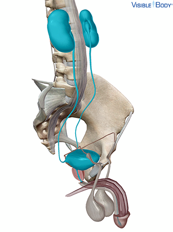
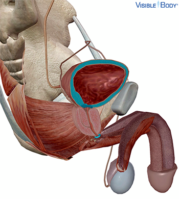
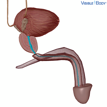
In men, the bladder sits in front of the rectum and above the prostate gland. The urinary system filters blood to remove waste through urine, which is voided through the urethra. In the male, the urethra extends through the prostate and penis.
System: Urinary
Region: Abdomen, Pelvis
Pathologies: Chronic kidney disease, diabetes insipidus, diabetes type 1, diabetes type 2, interstitial cystitis, kidney cysts, kidney failure, kidney stones, overactive bladder, rheumatoid arthritis, urinary incontinence, urinary tract infections, Wilms’ Tumor
Ren
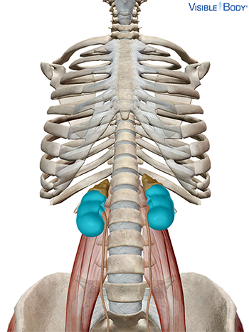
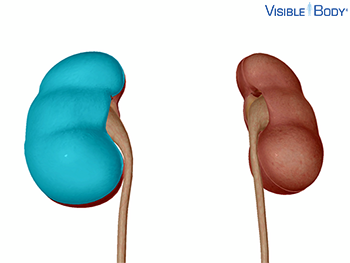
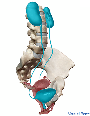
The kidneys are bean-shaped organs situated on the back of the abdominal wall, behind the peritoneum. The right kidney sits slightly lower than the left to accommodate the liver. The kidneys are composed of an external, lighter-colored cortex; an internal, darker medulla in which are found the renal pyramids; and a funnel-shaped structure called the renal pelvis. See it in 3D!
System: Urinary
Region: Abdomen
Function: The kidneys filter blood (supplied by the renal arteries) to remove unwanted substances. They also secrete waste into the urine.
Pathologies: Chronic kidney disease, diabetes insipidus, diabetes type 1, diabetes type 2, kidney cysts, kidney failure, kidney stones, rheumatoid arthritis, urinary tract infections, Wilms’ Tumor
Arteria renalis
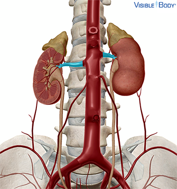
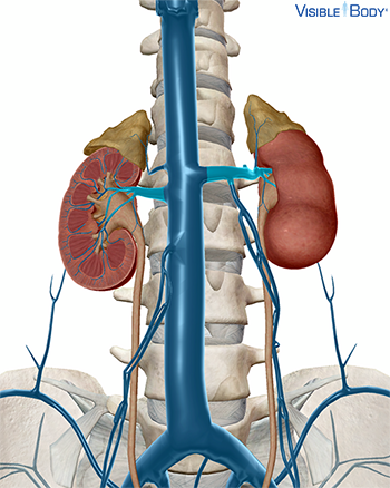
The renal arteries are wide trunks arising from the side of the abdominal aorta immediately below the superior mesenteric artery, between the first and second lumbar vertebrae.
System: Circulatory
Region: Abdomen
Function: Blood enters the kidneys through renal arteries. These arteries branch into tiny capillaries that interact with urinary structures inside the kidneys (namely the nephrons).
Pathologies: Aortic aneurysm, arteriovenous malformation, diabetes type 1, diabetes type 2
Vena renalis
These large vessels pass in front of the renal arteries and each splits into an anterior and posterior branch upon entering the kidneys.
System: Circulatory
Region: Abdomen
Function: The renal veins on the right and left sides of the abdomen drain the kidneys.
Pathologies: Arteriovenous malformations, diabetes type 1, diabetes type 2
Capsula fibrosa renis
Each kidney is surrounded and supported by three layers of tissue: the renal capsule, the adipose capsule, and the renal fascia.
System: Urinary
Region: Abdomen
Function: The transparent renal capsule is the innermost layer, composed of fibrous connective tissue that prevents trauma and infection from reaching the kidney and helps it maintain its shape.
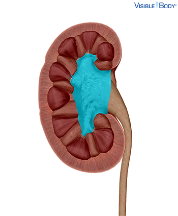
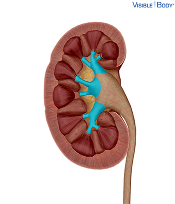
Sinus renalis
The renal sinus is the central cavity inside the kidney, located behind the hilum. It contains the renal pelvis, renal calyces, blood vessels, nerves, and adipose tissue (fat).
System: Urinary
Region: Abdomen
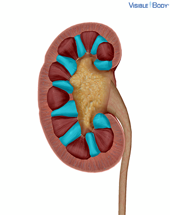
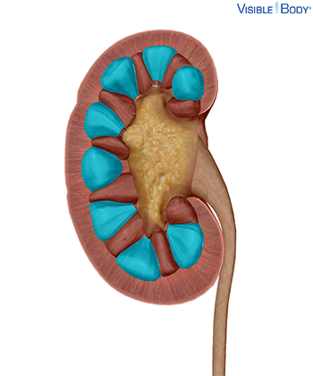
Columnae renales
The soft and granular renal cortex arches over the bases of the renal pyramids and dips in between them, forming the renal columns.
System: Urinary
Region: Abdomen
Function: These columns, composed of blood vessels, urinary tubes, and fibrous material, help anchor the renal cortex.
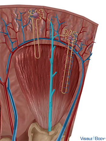
Pyramides renales
The renal pyramids (r, l) are a series of striated conical masses of tubules located within the medulla of the kidneys (r, l). Most of the renal pyramids’ mass is nephrons, the blood-filtering and urine-creating structures of the urinary system.
System: Urinary
Region: Abdomen
Nephroneum
Each kidney contains over 1 million tiny structures called nephrons. The nephrons are located partly in the cortex and partly inside the renal pyramids, where the nephron tubules make up most of the pyramid mass.
System: Urinary
Region: Abdomen
Function: Nephrons perform the primary function of the kidneys: regulating the concentration of water and other substances in the body. They filter the blood, reabsorb what the body needs, and excrete the rest as urine.
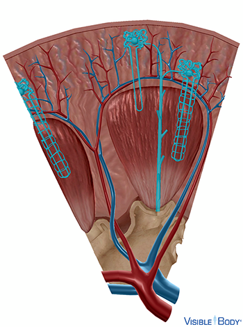
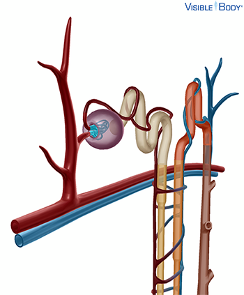
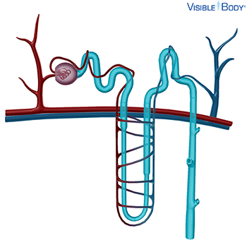
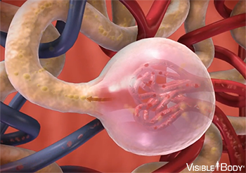
Glomerulus renalis
Each nephron has a glomerulus, the site of blood filtration. The glomerulus is a network of capillaries surrounded by a cuplike structure, the glomerular capsule (or Bowman’s capsule).
System: Urinary, Circulatory
Region: Abdomen
Function: As blood flows through the glomerulus, blood pressure pushes water and solutes from the capillaries into the capsule through a filtration membrane. This glomerular filtration begins the urine formation process.
Tubuli renales
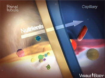
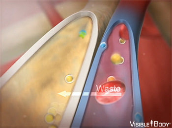
When the filtrate exits the glomerulus, it flows into a duct in the nephron called the renal tubule. As it moves, the needed substances and some water are reabsorbed through the tube wall into adjacent capillaries. This reabsorption of vital nutrients from the filtrate is the second step in urine creation.
System: Urinary
Region: Abdomen
Tubulus renalis colligens
Several nephrons supply filtrate to each collecting duct, and these ducts join together near the renal pelvis, draining urine through the renal pyramid papillae into the minor calyces. Filtrate that reaches the collecting duct is called urine.
System: Urinary
Region: Abdomen
Blood from the branches of the renal artery is filtered by nephrons in the renal pyramids. As blood flows through the glomerulus, blood pressure forces it against a specialized layer of cells that surrounds the capillaries. The layer of cells blocks blood cells and proteins, but lets waste and water pass through the glomerulus and into the glomerular capsule. The filtrate then flows into the proximal convoluted tubule.
System: Urinary
Region: Abdomen
As the filtrate passes out of the glomerular capsule and through the renal tubule, substances like water, essential ions, glucose, amino acids, and proteins are reabsorbed into the body through cells along the tube wall.
System: Urinary
Region: Abdomen
The filtrate absorbed in the glomerulus flows through the renal tubule, where nutrients and water are reabsorbed into capillaries. At the same time, waste ions and hydrogen ions pass from the capillaries into the renal tubule.
System: Urinary
Region: Abdomen
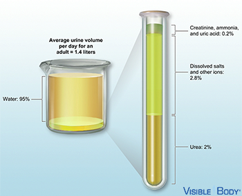
Urine is about 95% water and 5% waste products. Nitrogenous wastes excreted in urine include urea, creatinine, ammonia, and uric acid. Ions such as sodium, potassium, hydrogen, and calcium are also excreted.
System: Urinary
Region: Abdomen, Pelvis
Calices renales minores, calices renales majores
Before passing through the renal pelvis, urine passes through the minor and major calyces, cup-shaped tubes that surround the renal papillae. At the apex of the renal pyramids, urine passes through a renal papilla into a minor calyx, which converges with other minor calyces to form a major calyx. From the major calyces, urine drains into the renal pelvis.
System: Urinary
Region: Abdomen
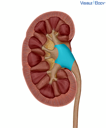
Pelvis renalis
The renal pelvis (r, l) is a funnel-shaped structure that begins within the kidney (r, l) and then protrudes from the kidney to join the ureter (r, l).
System: Urinary
Region: Abdomen
Function: As part of the urinary system, the renal pelvis functions as an excretory channel for the kidneys, funneling urine from the kidneys into the ureters.
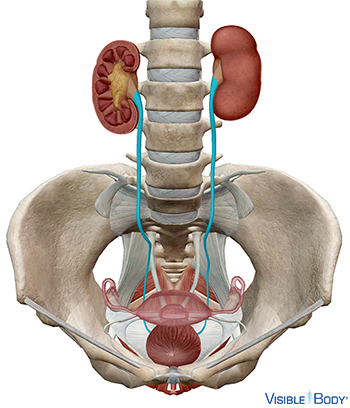
Urine drains from the renal pelvis of each kidney into the ureters. The ureters are long, thin tubes made of smooth muscle. In adults, the ureters are 25–30 cm long, about the length of a 12-inch ruler.
System: Urinary
Region: Abdomen, Pelvis
Function: Contractions of the smooth muscle push urine down through the ureters and into the bladder.
Pathologies: Urinary tract infections
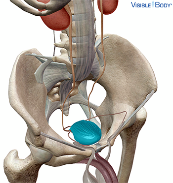
Vesica urinaria
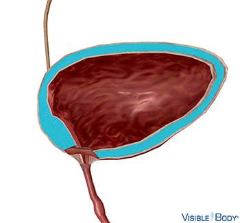
Urine flows through the ureters into the urinary bladder. The urinary bladder is an extremely elastic musculomembranous sac in the urinary system that serves as a reservoir for urine. The bladder is shaped like a pyramid when empty. It becomes more oval as it fills with urine and expands. A smooth muscle called the detrusor surrounds the bladder, and folds called rugae line the interior wall. These structures give the bladder elasticity and allow it to expand. See it in 3D!
System: Urinary
Region: Pelvis
Pathologies: Interstitial cystitis, kidney stones, overactive bladder, urinary incontinence, urinary tract infections
Musculus detrusor vesicae urinariae
At about 200 ml of urine, the detrusor muscle begins to contract and the internal urethral sphincter muscle begins to relax. This sends signals through the nervous system and creates the “urge” to urinate. If this urge is ignored, continence may be threatened. At about 500 ml, detrusor muscle contractions begin to force open the internal urethral sphincter. Unless the external urethral sphincter is powerful enough to prevent it, micturition (urination) will occur involuntarily.
System: Muscular, Urinary
Region: Pelvis
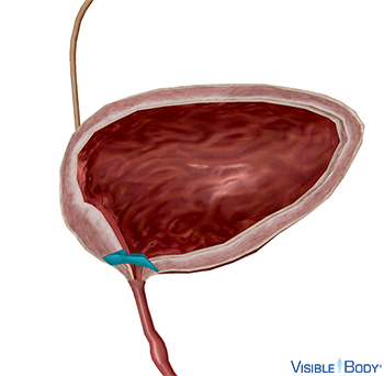
Musculus sphincter urethrae internus
The internal sphincter is involuntary. It surrounds the opening of the bladder to the urethra and relaxes to allow urine to pass.
System: Muscular, Urinary
Region: Pelvis
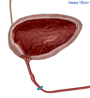
Musculus sphincter urethrae externus
The external sphincter is voluntary. It surrounds the urethra outside the bladder and must be relaxed for urination to occur.
System: Muscular, Urinary
Region: Pelvis
Micturition, or urination, is the act of emptying the bladder. When the bladder is full of urine, stretch receptors in the bladder wall trigger the micturition reflex. The detrusor muscle that surrounds the bladder contracts. The internal urethral sphincter relaxes, allowing for urine to pass out of the bladder into the urethra. Both of these reactions are involuntary. The external urethral sphincter is voluntary. It must be relaxed for urine to flow through the urethra and outside the body.
System: Muscular, Nervous, Urinary
Region: Pelvis
Urethra
In females, the urethra is narrow and about 4 cm long, significantly shorter than in males. It extends from the bladder neck to the external urethral orifice in the vestibule of the vagina.
System: Urinary
Region: Pelvis
Function: Urine produced in the kidneys passes through the ureters, collects in the bladder, and is then excreted through the urethra.
Pathologies: Urinary tract infections
Urethra
In males, the urethra is about 17.5–20 cm, four or five times as long as in females. The male urethra is divided into three sections: the prostatic urethra (the widest portion), the membranous urethra (the narrowest portion), and the spongy urethra (the longest portion). It extends from the bladder neck through the prostate and the penis to the external urethral orifice. See it in 3D!
System: Urinary
Region: Pelvis
Function: In men, both urine and semen pass out of the body through the urethra.
Pathologies: Urinary tract infections