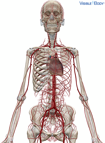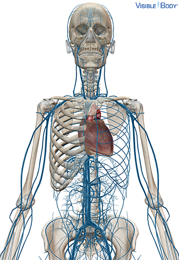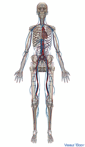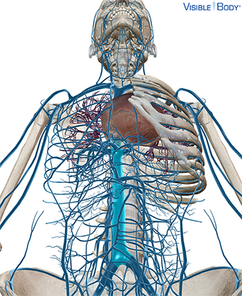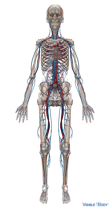
The circulatory system is composed of the heart and the blood vasculature: the arteries and the veins. See it in 3D!
System: Circulatory
Region: All
Function: Arteries carry blood away from the heart, and veins carry blood back to the heart. Circulating blood supplies cells throughout the body with oxygen and removes waste carbon dioxide.
Pathologies: Acute myeloid leukemia, anemia, angina, aortic aneurysm, aplastic anemia, arrhythmia, arteriovenous malformations, atherosclerosis, atrial fibrillation, blood clots, brain aneurysm, cardiomyopathy, carotid artery disease, childhood leukemia, chronic myeloid leukemia, congenital heart defects, coronary artery disease, deep vein thrombosis, diabetes type 1, diabetes type 2, diabetic foot, diabetic heart disease, giant cell arteritis, heart attack, heart failure, heart valve diseases, hemophilia, high blood pressure, Kawasaki Disease, low blood pressure, malaria, mitral valve prolapse, muscular dystrophy, pericardial disorders, peripheral arterial disease, Reynaud’s Disease, Rh incompatibility, rheumatoid arthritis, sickle cell anemia, stroke, thalassemia, transient ischemic attack, varicose veins, vasculitis
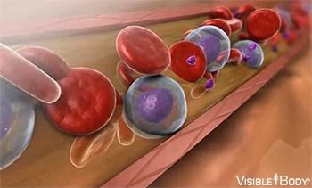
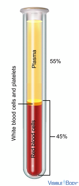
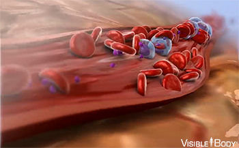
Blood is composed of 55% plasma and 45% “formed elements,” including red blood cells, white blood cells, and platelets. Because of these living cells suspended in the plasma, blood is considered a fluid connective tissue (not a fluid).
System: Circulatory
Region: All
Function: It transports oxygen and other essential substances throughout the body, fights sickness, and performs other vital functions.
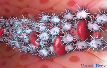
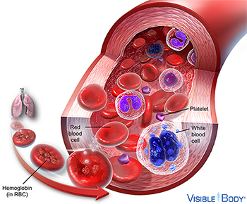
Cell fragments called platelets, or thrombocytes, make up about 2 percent of blood. See it in 3D!
System: Circulatory
Region: All
Function: Platelets clump and form a plug in the damaged area of a torn blood vessel to stop blood loss.
Red blood cells, also called erythrocytes, make up 40–45% of blood volume and function to transport oxygen from the lungs to the cells of the body. See it in 3D!
System: Circulatory
Region: All
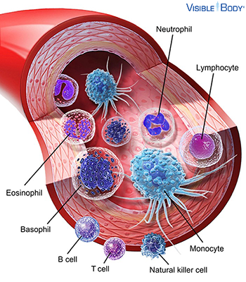
White blood cells, also called leukocytes, are the disease-fighting components of blood. They account for just 1% of circulating blood but multiply during infection or inflammation. See it in 3D!
System: Circulatory, Lymphatic (Immune)
Region: All
The largest component of blood is plasma, a yellowish liquid that is 90% water.
System: Circulatory
Region: All
Function: Plasma carries suspended blood cells and other substances.
Arteries transport blood away from the heart. In the systemic circulation arteries and their branches transport oxygenated blood and veins carry deoxygenated blood.
System: Circulatory
Region: All
Pathologies: Aortic aneurysm, arteriovenous malformations, diabetes type 1, diabetes type 2
Veins return blood back toward the heart. In the systemic circulation arteries and their branches transport oxygenated blood and veins carry deoxygenated blood. See it in 3D!
System: Circulatory
Region: All
Pathologies: Arteriovenous malformations, diabetes type 1, diabetes type 2
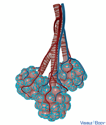
Capillaries surround body cells and tissues to deliver and absorb oxygen, nutrients, and other substances. The capillaries also connect the branches of arteries and to the branches of veins.
System: Circulatory
Region: All
Systemic circulation moves blood between the heart and the rest of the body. It sends oxygenated blood out to cells and returns deoxygenated blood to the heart.
System: Circulatory
Region: All
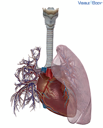
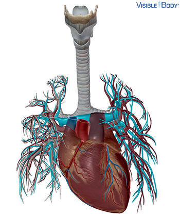
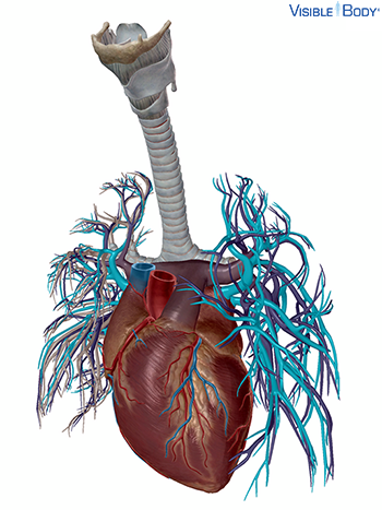
Pulmonary circulation moves blood between the heart and the lungs. It transports deoxygenated blood to the lungs to absorb oxygen and release carbon dioxide. The oxygenated blood then flows back to the heart.
System: Circulatory
Region: Thorax
Cor
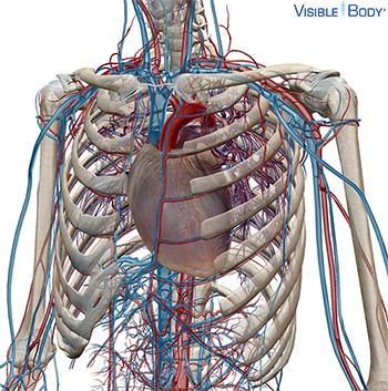
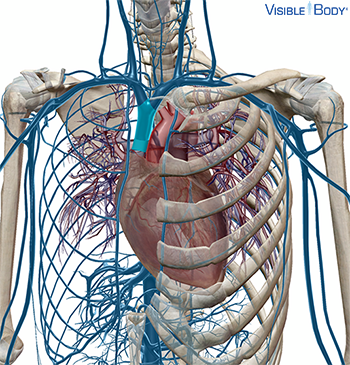
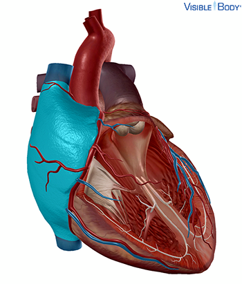
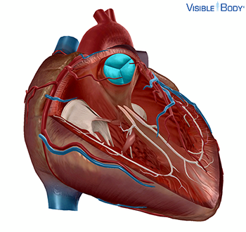
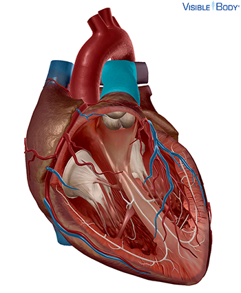
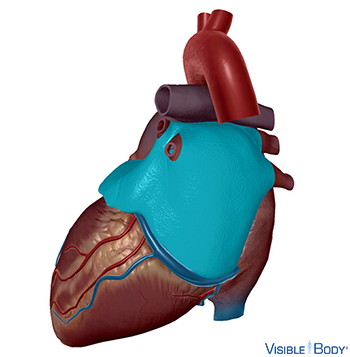
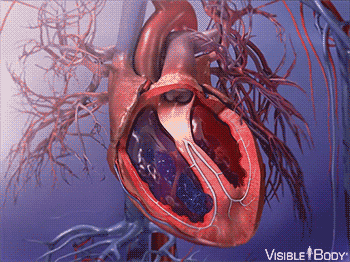
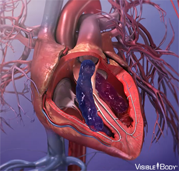
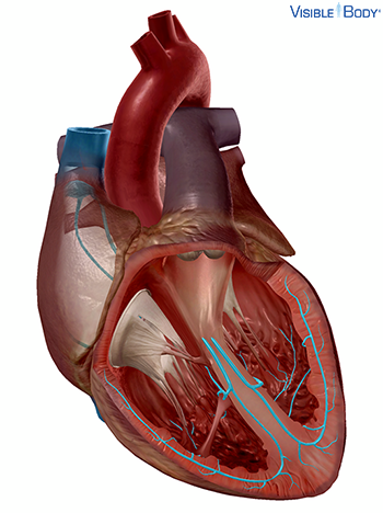
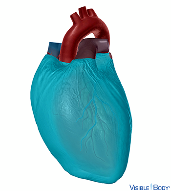
The heart is a hollow, muscular organ that pumps oxygenated blood throughout the body and deoxygenated blood to the lungs. The heart comprises four chambers enclosed by a heart wall. See it in 3D!
System: Circulatory
Region: Thorax
Pathologies: Angina, arrhythmia, atrial fibrillation, cardiomyopathy, congenital heart defects, coronary artery disease, diabetes type 1, diabetes type 2, diabetic heart disease, heart attack, heart failure, heart valve diseases, mitral valve prolapse, muscular dystrophy, pericardial disorders, rheumatoid arthritis
Pericardium
The pericardium is a fibroserous sac that contains the heart and the roots of the great vessels.
System: Circulatory
Region: Thorax
The muscular wall of the heart has three layers. The outermost layer is the epicardium (or visceral pericardium). The epicardium covers the heart, wraps around the roots of the great blood vessels, and adheres the heart wall to a protective sac. The middle layer is the myocardium. This strong muscle tissue powers the heart’s pumping action. The innermost layer, the endocardium, lines the interior structures of the heart.
System: Circulatory, Muscular
Region: Thorax
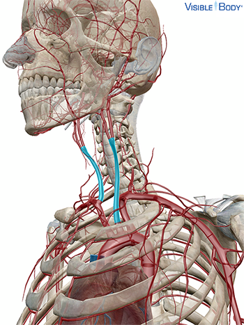
Vena cava superior
This large vessel is formed by the junction of the two brachiocephalic veins on either side of the root of the neck.
System: Circulatory
Region: Thorax
Function: The superior vena cava drains blood from the upper half of the body.
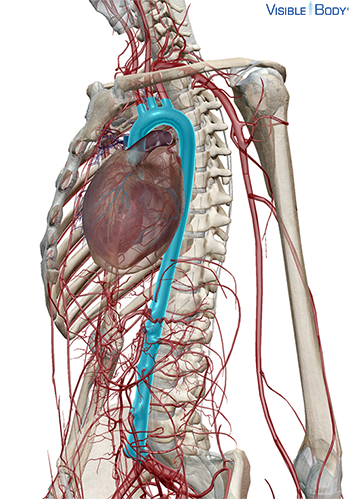
Vena cava inferior
This vessel ascends along the front of the vertebral column on the right side of the aorta. It perforates the diaphragm and passes behind the serous pericardium, opening into the right atrium at its base.
System: Circulatory
Region: Thorax, Abdomen
Function: The inferior vena cava returns blood to the heart from the abdominal walls and viscera below the diaphragm.
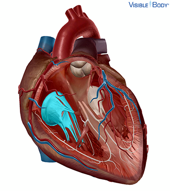
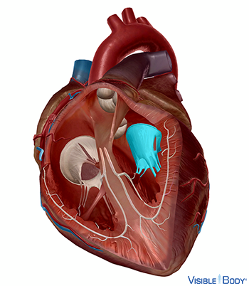
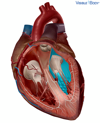
Atrium dextrum
The right atrium, one of the two upper chambers of the heart, receives deoxygenated blood from the vena cava (superior, inferior) and the coronary sinus, and empties into the right ventricle.
System: Circulatory
Region: Thorax
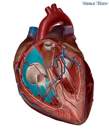
Valva atrioventricularis dextra (valva tricuspidalis)
The tricuspid valve, one of the two atrioventricular valves in the heart, controls blood flow from the right atrium into the right ventricle.
System: Circulatory
Region: Thorax
Function: The tricuspid valve plays a vital role in the cardiac cycle, preventing backflow and ensuring one-way blood flow through the heart. When the right ventricle contracts, the tricuspid valve is closed, preventing the passage of the blood back into the atria.
Ventriculus dexter
The right ventricle, one of the two lower chambers of the heart, is responsible for pumping deoxygenated blood into the pulmonary trunk.
System: Circulatory
Region: Thorax
Valva trunci pulmonalis
The pulmonary semilunar valve, one of the four heart valves, controls blood flow from the right ventricle into the pulmonary trunk.
System: Circulatory
Region: Thorax
Function: The pulmonary semilunar valve plays a role in the cardiac cycle, conveying deoxygenated blood from the heart into the lungs.
Truncus pulmonalis
The pulmonary trunk, a great vessel of the cardiovascular system, supports pulmonary circulation by carrying deoxygenated blood from the right ventricle of the heart into the lungs for gas exchange. At the aortic arch, the pulmonary trunk divides into the pulmonary arteries (r, l), which extend into the lungs. The trunk and its branching arteries are the only arteries in the body that carry deoxygenated blood.
System: Circulatory
Region: Thorax
Pathologies: Diabetes type 1, diabetes type 2
Arteriae pulmonales
The right and left pulmonary arteries stem from the pulmonary trunk, which connects at its base to the right ventricle of the heart. In the lungs, the pulmonary arteries branch into arterioles and then into networks of pulmonary capillaries. Gases are exchanged across the walls of these capillaries, and the oxygen content of the blood rises.
System: Circulatory
Region: Thorax
Function: Together, the pulmonary arteries convey deoxygenated blood from the right ventricle to the lungs.
Pathologies: Diabetes type 1, diabetes type 2
Venae pulmonales superiores, venae pulmonales inferiores
The pulmonary veins are formed by the joining of venules from pulmonary capillary beds in the lungs. They are four in number—two from each lung—and have no valves. The pulmonary veins are the only veins in the body that carry oxygenated blood.
System: Circulatory
Region: Thorax
Function: These veins return oxygenated blood from the lungs to the left atrium of the heart for distribution via systemic circulation to the rest of the body.
Pathologies: Diabetes type 1, diabetes type 2
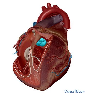
Atrium sinistrum
The left atrium, one of the two upper chambers of the heart, receives oxygenated blood from the pulmonary veins, and empties into the left ventricle.
System: Circulatory
Region: Thorax
Valva atrioventricularis sinistra (valva mitralis)
The mitral valve, one of the two atrioventricular valves in the heart, controls blood flow from the left atrium into the left ventricle.
System: Circulatory
Region: Thorax
Function: The mitral valve plays a role in the cardiac cycle, conveying oxygenated blood through the heart. When the left ventricle contracts, the mitral valve is closed; this action prevents the backflow of blood into the left atrium.
Ventriculus sinister
The left ventricle, one of the two lower chambers of the heart, is responsible for pumping oxygenated blood into the aorta.
System: Circulatory
Region: Thorax
Valva aortae
The aortic semilunar valve, one of four heart valves, regulates and supports the one-way flow of blood out of the left ventricle and into the aorta; the opening and closing of the aortic semilunar valve, a result of varying blood pressure, contributes to the cardiac cycle of the cardiovascular system.
System: Circulatory
Region: Thorax
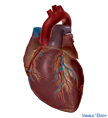
Aorta
The aorta, a great vessel of the cardiovascular system, is the largest artery in the body. It begins at its base at the left ventricle of the heart, where it receives blood from the left ventricle through the aortic valve. See it in 3D!
System: Circulatory
Region: Thorax
Arteria carotis communis
The left and right common carotid arteries are the principal arteries of supply to the head and neck. As the common carotids ascend in the neck, each divides into two branches at the level of the upper border of the thyroid cartilage.
System: Circulatory
Region: Thorax, Neck
Pathologies: Aortic aneurysm, arteriovenous malformations, atherosclerosis, brain aneurysm, carotid artery disease, diabetes type 1, diabetes type 2, stroke, transient ischemic attack
The cardiac cycle regulates both electrical and mechanical activities of the heart. An electrical impulse is sent, which results in a mechanical action. The cardiac cycle splits into two phases: systole and diastole. Ventricular contraction and constriction is known as systole, while relaxation and expansion of the ventricles is called diastole. Each contraction and relaxation is a heartbeat. The rate of contractions in a healthy heart is about 60 to 70 beats per minute when the body is at rest. During physical activity, the heart beats about 100–120 times per minute.
System: Circulatory
Region: Thorax
Ventricular contractions, called systole, force blood out of the heart through the pulmonary and aortic valves. This phase begins with the closure of the AV valves and ends with the closure of the semilunar valves.
System: Circulatory
Region: Thorax
Diastole occurs when blood flows from the atria to fill the ventricles. Diastole starts with the closure of the semilunar valves and ends with the closure of the AV valves.
System: Circulatory
Region: Thorax
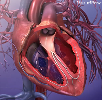
The conduction system of the heart is a system within the heart that is controlled by the autonomic nervous system and that delivers electrical impulses and motivates the rhythmic contractions of the heart. The pathways for the electrical impulses are formed by a series of bundles of specialized muscle fibers within the heart: sinoatrial node, atrioventricular node, atrioventricular bundle of His, left and right bundle branches, and Purkinje fibers. There are distinct steps to each electrical impulse; these result in the contraction of the heart’s upper chambers followed by the contraction of the heart’s lower chambers.
System: Circulatory
Region: Thorax
Blood pressure is the amount of force put on blood vessels. It is caused by the blood flow generated by the heart as it pumps, and the resistance that blood encounters as it moves through the enclosed vessel. The point of highest pressure, when the ventricles are contracting and pressure is highest in the arteries, is called systolic pressure. The point of lowest pressure, when the ventricles are relaxed and the semilunar valves are closed, is called diastolic pressure. The average systolic pressure is 120 millimeters of mercury. The average diastolic pressure is 70 to 80 millimeters of mercury.
System: Circulatory