


The nervous system must receive and process information about the world outside in order to react, communicate, and keep the body healthy and safe. Much of this information comes through the sensory organs: the eyes, ears, nose, tongue, and skin. Specialized cells and tissues within these organs receive raw stimuli and translate them into signals the nervous system can use. Nerves relay the signals to the brain, which interprets them as sight (vision), sound (hearing), smell (olfaction), taste (gustation), and touch (tactile perception).

The eyes sit in the orbits of the skull, protected by bone and fat. The white part of the eye is the sclera. It protects interior structures and surrounds a circular portal formed by the cornea, iris, and pupil. The cornea is transparent to allow light to enter the eye, and curved to direct it through the pupil behind it. The pupil is actually an opening in the colored disk of the iris. The iris dilates or constricts, adjusting how much light passes through the pupil and onto the lens. The curved lens then focuses the image onto the retina, the eye’s interior layer. The retina is a delicate membrane of nervous tissue containing photoreceptor cells. These cells, the rods and cones, translate light into nervous signals. The optic nerve carries the signals from the eye to the brain, which interprets them to form visual images.
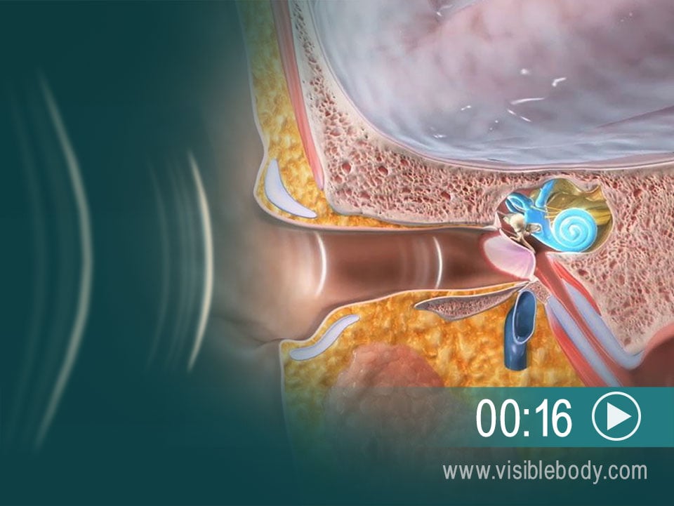
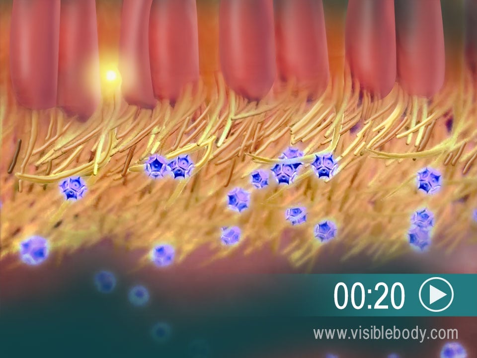
Music, laughter, car honks — all reach the ears as sound waves in the air. The outer ear funnels the waves down the ear canal (the external acoustic meatus) to the tympanic membrane (the “ear drum”). The sound waves beat against the tympanic membrane, creating mechanical vibrations in the membrane. The tympanic membrane transfers these vibrations to three small bones, known as auditory ossicles, found in the air-filled cavity of the middle ear. These bones – the malleus, incus, and stapes – carry the vibrations and knock against the opening to the inner ear. The inner ear consists of fluid-filled canals, including the spiral-shaped cochlea. As the ossicles pound away, specialized hair cells in the cochlea detect pressure waves in the fluid. They activate nervous receptors, sending signals through the cochlear nerve toward the brain, which interprets the signals as sounds.

Skin consists of three major tissue layers: the outer epidermis, middle dermis, and inner hypodermis. Specialized receptor cells within these layers detect tactile sensations and relay signals through peripheral nerves toward the brain. The presence and location of the different types of receptors make certain body parts more sensitive. Merkel cells, for example, are found in the lower epidermis of lips, hands, and external genitalia. Meissner corpuscles are found in the upper dermis of hairless skin — fingertips, nipples, the soles of the feet. Both of these receptors detect touch, pressure, and vibration. Other touch receptors include Pacinian corpuscles, which also register pressure and vibration, and the free endings of specialized nerves that feel pain, itch, and tickle.
The sense of smell is called olfaction. It starts with specialized nerve receptors located on hairlike cilia in the epithelium at the top of the nasal cavity. When we sniff or inhale through the nose, some chemicals in the air bind to these receptors. That triggers a signal that travels up a nerve fiber, through the epithelium and the skull bone above, to the olfactory bulbs. The olfactory bulbs contain neuron cell bodies that transmit information along the cranial nerves, which are extensions of the olfactory bulbs. They send the signal down the olfactory nerves, toward the olfactory area of the cerebral cortex.

What are all those small bumps on the top of the tongue? They’re called papillae. Many of them, including circumvallate papillae and fungiform papillae, contain taste buds. When we eat, chemicals from food enter the papillae and reach the taste buds. These chemicals (or tastants) stimulate specialized gustatory cells inside the taste buds, activating nervous receptors. The receptors send signals to fibers of the facial, glossopharyngeal, and vagus nerves. Those nerves carry the signals to the medulla oblongata, which relays them to the thalamus and cerebral cortex of the brain.
