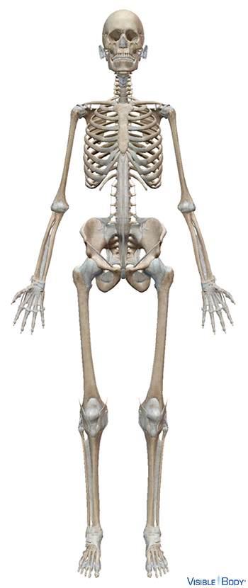
The bones of the human skeletal system are divided into an axial region and an appendicular region. See it in 3D!
System: Skeletal
Region: All
Function: The skeleton is the framework that provides structure to the rest of the body and facilitates movement.
Pathologies: Dislocations, fractures, herniated disc, infectious arthritis, osteoarthritis, osteoporosis
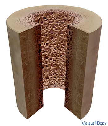
There are five types of bones in the skeleton: flat, long, short, irregular, and sesamoid. Bones are composed of an exterior layer of compact bone tissue that surrounds the internal spongy bone tissue.
System: Skeletal
Region: All
Function: Bones of the appendicular skeleton support and facilitate movement, while bones of the axial skeleton protect internal organs. The skeleton also stores minerals such as calcium, and produces red blood cells.
Pathologies: Osteoarthritis, osteogenesis imperfecta, osteonecrosis, osteoporosis, Paget’s Disease of Bone
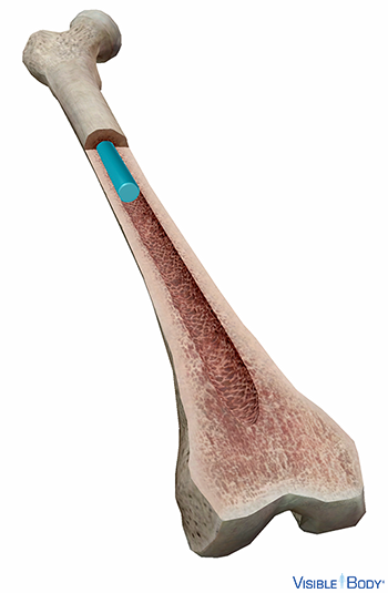
Bone marrow fills the cylindrical cavities in the bodies of long bones and occupies the spaces inside spongy bone. It also extends into the larger bony canals (central canals) that contain blood vessels. See it in 3D!
System: Skeletal, Immune
Region: Long and spongy bones of skeleton
Function: Yellow marrow is found in the central cavities of long bones and consists mostly of fat. Red marrow is found in the medullary cavities of flat and short bones, the articular ends of long bones, the bodies of vertebrae, the spongy bone of the cranium, the sternum, the scapulae, and the ribs. Red marrow consists mostly of hematopoietic tissue, with a small amount of fat. It is essential for red blood cell production and contains more marrow cells, or myelocytes, than yellow marrow.
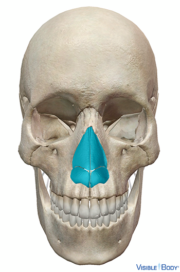
Cartilage is more flexible than bone but stiffer than muscle. See it in 3D!
System: Skeletal
Region: All
Function: Cartilage gives structure to the larynx and nose. It is also found between the vertebrae and at the ends of bones like the femur.
Pathologies: Osteoarthritis
Ligaments are bands of dense connective tissue that are key to the function of joints. See it in 3D!
System: Skeletal
Region: All
Function: Synovial joints are often supported and reinforced by surrounding ligaments, which limit movement to prevent injury. A syndesmosis is a joint in which a ligament connects two bones, allowing for a little movement (amphiarthroses).
Pathologies: Sprains
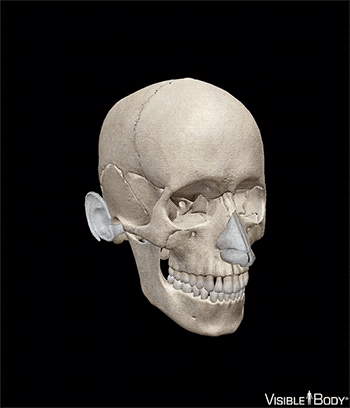
The skull comprises most of the bones of the head region. It is divided into two parts: the eight bones of the neurocranium and the fourteen bones of the facial skeleton. See it in 3D!
System: Skeletal (Axial)
Region: Head
Function: The cranium (neurocranium) includes the bones of the skull that enclose and protect the brain. The facial bones include all the bones located in the anterior portion of the skull that create the facial skeleton. These bones give shape to the face; support the primary organs of smell, taste, and sight; and are shaped and joined in a manner that provides openings for the respiratory and digestive systems.
Pathologies: Fractures
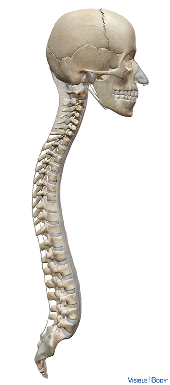
The vertebral column is a flexible column formed by a series of vertebrae that extend from the base of the skull to the pelvis. See it in 3D!
System: Skeletal (Axial)
Region: Neck, Thorax, Back
Function: The vertebral column articulates with the vertebrosternal ribs and false ribs, supports the skull and trunk, and protects the spinal cord, which passes through the vertebral foramina.
Pathologies: Scoliosis, spinal stenosis
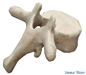
Vertebra cervicalis, vertebra thoracica, vertebra lumbalis
The cervical vertebrae are the first seven (C01-C07) of the 33 vertebrae of the vertebral column. The thoracic vertebrae (T01-T12) are the 8th through 19th of the 33 vertebrae of the vertebral column. The lumbar vertebrae (L01-L05) are the 20th through 24th of the 33 vertebrae of the vertebral column. See it in 3D!
System: Skeletal (Axial)
Region: Neck, Thorax, Back
Function: The disc-like body of the vertebrae is weight-bearing and its upper and lower surfaces give attachment to the intervertebral discs (intervertebral fibrocartilages).
Pathologies: Ankylosing spondylitis, bulging disc, cervical radiculopathy, cervical spondylosis, herniated disc
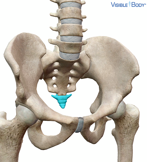
Os coccygis
The coccyx (tailbone) is the terminal portion of the vertebral column and forms part of the posterior wall of the pelvic cavity. See it in 3D!
System: Skeletal (Axial)
Region: Pelvis
Function: The anterior surface of the coccyx provides attachment for the anterior sacrococcygeal ligament and the levator ani and supports part of the rectum. The posterior surface, at the base, articulates with the sacrum by a fibrocartilage joint.
Pathologies: Ankylosing spondylitis, herniated disc
Os sacrum
The sacrum includes the sacral vertebrae (S01-S05); these are the 25th through 29th of the 33 vertebrae of the vertebral column.
System: Skeletal (Axial)
Region: Pelvis
Function: The vertebral canal runs throughout the greater part of the bone, forming a passage for the sacral nerves, and its walls are perforated by the anterior and posterior sacral foramina, through which these nerves exit.
Pathologies:, Ankylosing spondylitis, herniated disc
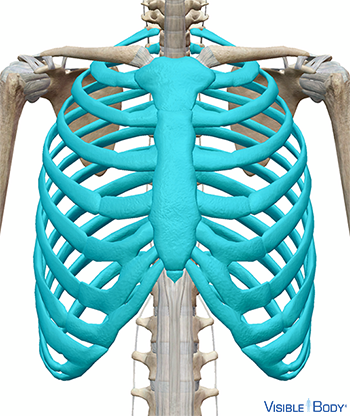
The thoracic cage is a flexible framework of bones and cartilages in the axial skeleton. See it in 3D!
System: Skeletal (Axial)
Region: Thorax
Function: Contain and protect the principal organs of respiration and circulation.
Pathologies: Fractures
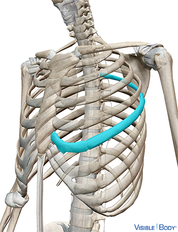
Costa vera, costa spuria
The vertebrosternal ribs (true ribs) are the first seven pairs (01-07) of ribs that form the thoracic cage. The false ribs (08-12) are the five inferior pairs of ribs that form part of the thoracic cage and give it flexibility. See it in 3D!
System: Skeletal (Axial)
Region: Thorax
Function: The vertebrosternal ribs provide support and flexibility to the thoracic cage.
Pathologies: Dislocations, fractures, osteogenesis imperfecta
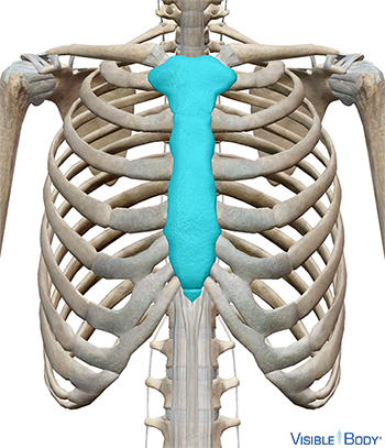
Corpus sterni, manubrium sterni, processus xiphoideus
The sternum is an elongated, flattened bone that forms the middle portion of the anterior wall of the thoracic cage and serves as an attachment for several muscles.
System: Skeletal (Axial)
Region: Thorax
Function: Its superior end supports the clavicles and its margins articulate with the costal cartilages of the first seven pairs of vertebrosternal ribs (true ribs).
Pathologies: Fractures
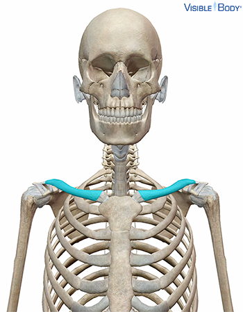
Clavicula
The clavicle (r, l) makes up the anterior portion of the shoulder girdle (pectoral girdle), the part of the appendicular skeleton that connects the bones of the upper limbs to the axial skeleton. See it in 3D!
System: Skeletal (Appendicular)
Region: Shoulder
Function: The clavicle is long and doubly curved and articulates medially with the cartilage of the first rib and the manubrium of the sternum and laterally with the acromion of the scapula. The clavicle’s upper surface is flat and rough and provides attachments for the deltoids anteriorly and the trapezius posteriorly.
Pathologies: Dislocations, fractures
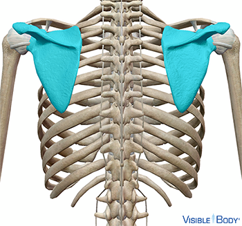
Scapula
The two scapulas (r, l) form the posterior portion of the shoulder girdle (pectoral girdle), the part of the appendicular skeleton that connects the bones of the upper limbs to the axial skeleton. See it in 3D!
System: Skeletal (Appendicular)
Region: Shoulder
Function: Laterally, the scapula articulates with the humerus via a ball and socket joint that provides extensive mobility to the shoulder joint. The medial border of the scapula gives attachment to a portion of the trapezius and articulates with the lateral end of the clavicle.
Pathologies: Dislocations
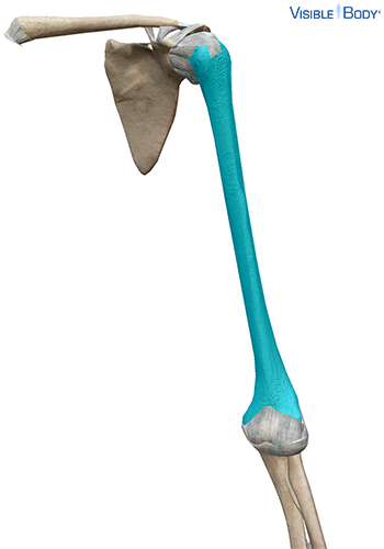
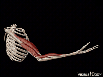
Humerus
The humerus is the longest and largest bone of the upper limb. See it in 3D!
System: Skeletal (Appendicular)
Region: Upper Limb
Function: Articulates with the radius and the ulna of each arm, forms part of the elbow, provides attachments for several shoulder and arm muscles.
Pathologies: Dislocations, fractures
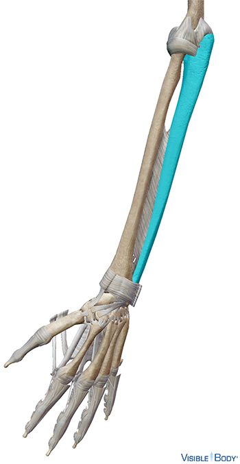
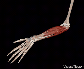
Radius
The radius (r, l) is a long, thin bone of the forearm (antebrachium)—a segment of the upper limb of the appendicular skeleton.
System: Skeletal (Appendicular)
Region: Upper Limb
Function: Articulates with the radius and the ulna of each arm, forms part of the elbow, provides attachments for several shoulder and arm muscles.
Pathologies: Dislocations, fractures
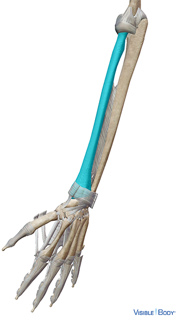
Ulna
The ulna (r, l) is a long, thin bone of the forearm (antebrachium)—a segment of the upper limb of the appendicular skeleton.
System: Skeletal (Appendicular)
Region: Upper Limb
Function: The ulna is parallel and medial to the radius. Together they provide rotational movement (supination and pronation) of the forearm.
Pathologies: Dislocations, fractures
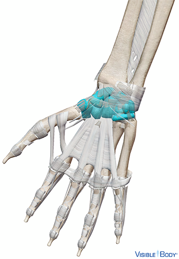
The eight carpals (r, l), carpus (wrist) bones of the hand, are arranged in two irregular rows.
System: Skeletal (Appendicular)
Region: Upper Limb (Hand)
Function: The carpals articulate with the bones of the metacarpals (palm) and the radius and the ulna of the forearm in such a way as to facilitate great mobility of the wrist and hand and to allow powerful use of the extensors and flexors of the forearm.
Pathologies: Dislocations, fractures
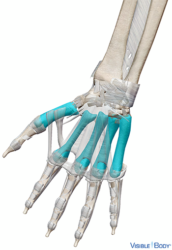
Os metacarpi
The metacarpals (r, l) are the bones of the middle part (palm) of the hand, which constitute the distal portions of the appendicular skeleton’s upper limbs.
System: Skeletal (Appendicular)
Region: Upper Limb (Hand)
Function: The medial and lateral surfaces are concave, for the attachment of the interosseous muscles.
Pathologies: Dislocations, fractures
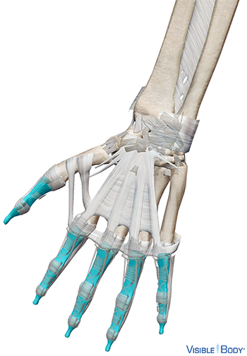
Phalanx proximalis, phalanx media, phalanx distalis
The phalanges of the hand (r, l), one of three regions of the hand, comprise the bones of the five digits of the hands (fingers).
System: Skeletal (Appendicular)
Region: Upper Limb (Hand)
Function: The first digit is adapted to oppose the movements of digits 2 through 5, thereby providing the ability to grasp and manipulate objects.
Pathologies: Dislocations, fractures
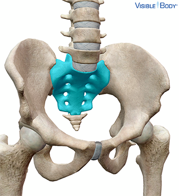
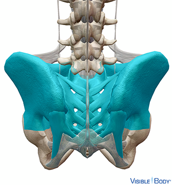
The pelvic girdle of the skeletal system, a ring of bones attached to the vertebral column, connect the bones of the lower limbs to the axial skeleton.
System: Skeletal (Appendicular)
Region: Pelvis, Hip
Function: Along with the coccyx and sacrum, the pelvic girdle forms the walls of the pelvic cavity, which protects some of the reproductive organs.
Pathologies: Dislocations, fractures
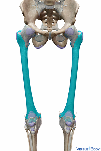
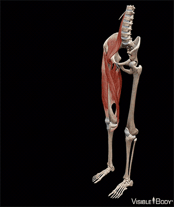
Os femoris
The femur (r, l) is the longest bone in the body and is the only bone located in the thigh of the upper leg. See it in 3D!
System: Skeletal (Appendicular)
Region: Lower Limb
Function: Adapted for weight bearing and mobility, the femur articulates with the pelvis at a ball and socket joint that provides the full range of motion required for walking and running.
Pathologies: Dislocations, fractures
Patella
The patella (r, l), located between the thigh and lower leg, is a flat, triangular bone that articulates with the patellar surface of the femur and protects the anterior surface of the knee. See it in 3D!
System: Skeletal (Appendicular)
Region: Lower Limb
Function: The superior and inferior surfaces provide an attachment point for the patellar ligament, a continuation of the common tendon of the quadriceps femoris.
Pathologies: Dislocations, fractures
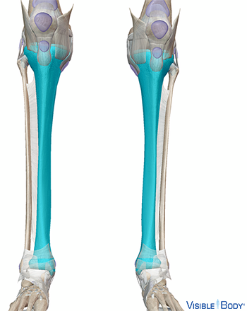
Tibia
The tibia (r, l) is the larger of the two parallel bones in the lower leg. See it in 3D!
System: Skeletal (Appendicular)
Region: Lower Limb
Function: The proximal end of the tibia is large, with a pair of condyles and two facets for articulation with the femur and ligaments of the knee. The body of the tibia (shaft) is adapted for weight bearing. The distal end of the tibia articulates with the fibula and the bones of the foot.
Pathologies: Dislocations, fractures
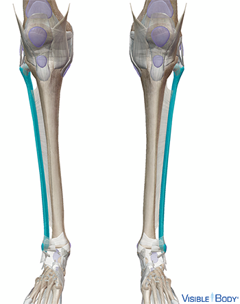
Fibula
The fibula (r, l) is the smaller of the two parallel bones in the lower leg.
System: Skeletal (Appendicular)
Region: Lower Limb
Function: The body of the fibula (shaft) is long, slender, and has four surfaces that provide attachments for various muscles and ligaments. The distal end projects below the tibia, forms the lateral part of the ankle, and provides attachment points for ligaments of the foot.
Pathologies: Dislocations, fractures
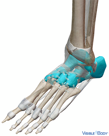
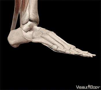
Ossa tarsalia
The tarsus (r, l), one of the three regions of the foot, includes seven tarsal bones that form the ankle, the heel, the transverse arch, and the medial part of the longitudinal arch of the foot.
System: Skeletal (Appendicular)
Region: Lower Limb (Foot)
Function: The bones of the foot articulate with the fibula and tibia at the upper surface of the talus where ligaments help form the ankle’s hinge joint.
Pathologies: Dislocations, fractures
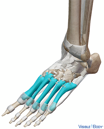
Ossa metatarsalia
The metatarsus (r, l), one of the three regions of the foot, comprises five metatarsal bones that form the sole and instep; the bones are numbered from the medial side (1 through 5).
System: Skeletal (Appendicular)
Region: Lower Limb (Foot)
Function: Each metatarsal bone consists of a tapered body (shaft) and two extremities. The wedge-shaped posterior extremity articulates proximally with one or more tarsals and with the contiguous metatarsal bones; its dorsal and plantar surfaces are rough for the attachment of ligaments.
Pathologies: Dislocations, fractures
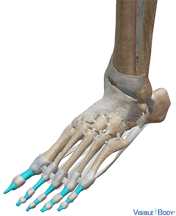
Phalanx proximalis, phalanx media, phalanx distalis
The metatarsus (r, l), one of the three regions of the foot, comprises five metatarsal bones that form the sole and instep; the bones are numbered from the medial side (1 through 5).
System: Skeletal (Appendicular)
Region: Lower Limb (Foot)
Function: The phalanges of the distal row resemble those of the fingers, but are smaller and are flatter; each has a broad base for articulation with the corresponding bone of the middle row and an expanded distal extremity for support of the nail and the end of the toe.
Pathologies: Dislocations, fractures
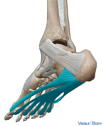
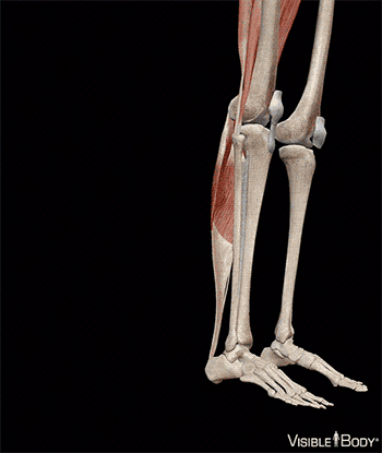
The plantar fascia (r, l), or plantar aponeurosis, is a long, thin fibrous band that extends from the heel to the digits on the plantar surface of the foot. See it in 3D!
System: Skeletal
Region: Lower Limb (Foot)
Function: The plantar fascia is of great strength and supports the arch.
Pathologies: Plantar fasciitis
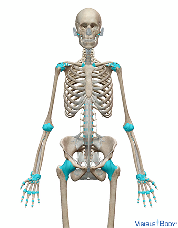
Joints throughout the body hold the skeleton together and support movement. Joints are categorized by the range of movement provided. See it in 3D!
System: Skeletal
Region: All
Pathologies: Infectious arthritis, osteoarthritis, sprains
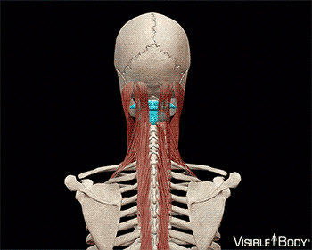
The atlas, along with the axis, articulates with and supports the skull to provide a pivot joint that allows for the great range of motion of the neck.
System: Skeletal
Region: Head and Neck
Type: Pivot
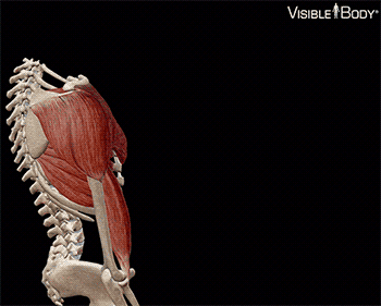
The ligaments of the shoulder girdle (r, l) include the acromioclavicular, coracoacromial, sternoclavicular, and superior transverse scapular ligaments. These ligaments connect the clavicle and the scapula of each shoulder girdle and function to support proper movement of the shoulder joint. See it in 3D!
System: Skeletal (Appendicular)
Region: Shoulder
Type: Ball and socket
Pathologies: Adhesive capsulitis (damaged articular capsule), labral tear (partially torn glenoid labrum)
The elbow joint, a hinge synovial joint, is enclosed by a thin articular capsule that allows the elbow to flex and extend freely. Thickened portions of the articular capsule form ligaments that support and stabilize the elbow joint.
System: Skeletal (Appendicular)
Region: Upper Limb
Type: Hinge
Pathologies: Lateral epicondylitis (tennis elbow), medial epicondylitis
The wrist is composed of eight carpal bones and numerous ligaments that keep these bones in place. These ligaments connect the bones of the wrist proximally to the radius and ulna and distally to the five metacarpals, allowing for stable movement of the wrist joint.
System: Skeletal (Appendicular)
Region: Upper Limb
Type: Condyloid
Pathologies: Carpal tunnel syndrome, wrist sprain
At the sacroiliac joints, the ilium articulates with the sacrum. Ligaments of the pelvis reinforce the articulations between the axial skeleton and the pelvic girdle.
System: Skeletal (Appendicular)
Region: Hip, Pelvis
Type: Gliding
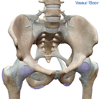
The ligaments of the hip connect the pelvis to the femur and support and stabilize the hip joint, which is a ball and socket joint. See it in 3D!
System: Skeletal (Appendicular)
Region: Hip
Type: Ball and socket
Pathologies: Hip labral tear, osteoarthritis
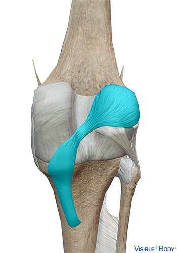
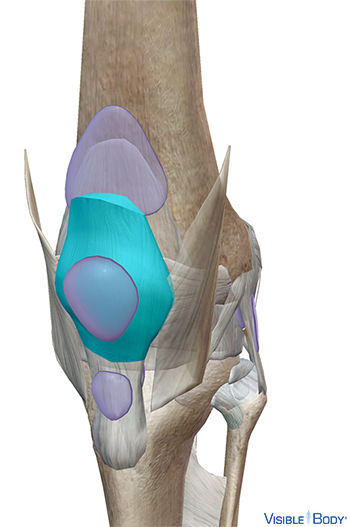
Together, the medial (r, l) and lateral (r, l) sections of the meniscus form a flat, curved pad of fibrocartilage that dissipates pressure and reduces friction in the knee joint.
System: Skeletal (Appendicular)
Region: Lower Limb
Type: Hinge
Pathologies: Knee sprain, torn meniscus, partially torn ACL
The bones of the foot articulate with the fibula and tibia at the upper surface of the talus where ligaments help form the ankle’s hinge joint. See it in 3D!
System: Skeletal (Appendicular)
Region: Lower Limb (Foot)
Type: Hinge
Pathologies: Ankle sprain