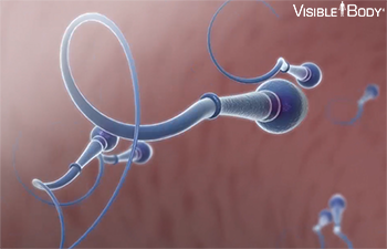
Gametes, the male and female sex cells, are produced through meiosis in the ovaries and testes.
System: Reproductive
Region: Pelvis
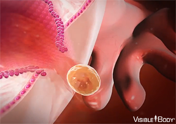
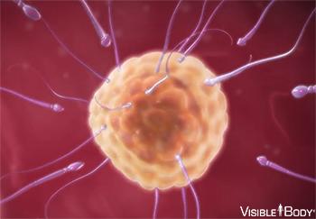
Function: The sperm and the egg are gametes. They each contain half the genetic information necessary for reproduction. When a sperm cell penetrates and fertilizes an egg, that genetic information combines.
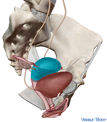
The ovaries produce secondary oocytes—the female sex cells. Each month one secondary oocyte is released into the uterine tube. If the oocyte is fertilized, it implants in the uterus.
System: Reproductive
Region: Pelvis
Ovarium
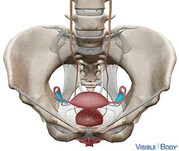
The ovaries are two almond-shaped structures that sit on either side of the uterus, connected to the uterine tubes. See it in 3D!
System: Endocrine, Reproductive
Region: Pelvis
Function: They produce oocytes (egg cells), as well as estrogen, progesterone, and other hormones.
Pathologies: Infertility, ovarian cysts, pelvic inflammatory disease, polycystic ovary syndrome, premature ovarian failure
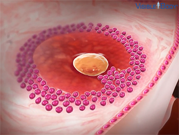
Egg cell production, or oogenesis, begins with the primordial follicles. As girls reach puberty, each ovary contains thousands of these follicles, and each follicle contains a primary oocyte. When follicles mature, some primary oocytes become secondary oocytes. By the time of ovulation, there is only one mature follicle remaining. The rest of the follicles deteriorate. During ovulation (about once a month), the dominant follicle bursts and releases its secondary oocyte. The oocyte travels into the uterine tube, where it can be fertilized.
System: Reproductive
Region: Pelvis
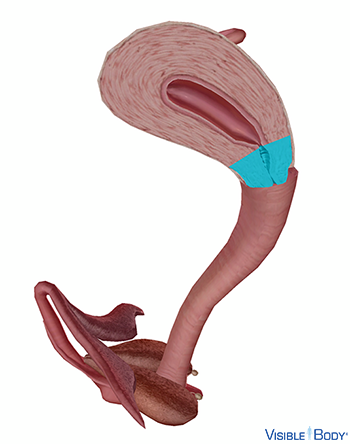
Tuba uterina
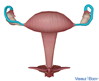
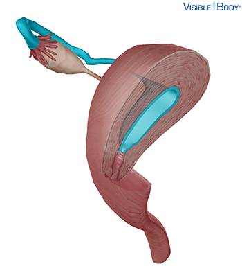
The uterine tubes (also called Fallopian tubes or oviducts) connect the ovaries to the uterus. Each uterine tube can be divided into three parts: The infundibulum is open to the abdomen. A constricted section called the isthmus connects with the uterus. Finally, an intermediate and dilated portion, the ampulla, curves over the ovary.
System: Reproductive
Region: Pelvis
Function: Egg fertilization usually occurs in the ampulla. The eggs then travel through the isthmus into the uterus.
Pathologies: Ectopic pregnancy, infertility
Uterus
The uterus is a pear-shaped organ located in the pelvic cavity between the bladder and the rectum. It is a hollow organ with thick, muscular walls. See it in 3D!
System: Reproductive
Region: Pelvis
Function: During menstruation, the inner lining of the uterus is shed. When a woman becomes pregnant, however, the fertilized egg embeds itself in the uterine wall and menstruation is prevented. The uterus expands dramatically as the egg develops into an embryo and then a growing fetus.
Pathologies: Endometriosis, gonorrhea, infertility, pelvic inflammatory disease, uterine fibroids
Women of childbearing age go through a cycle about every 28 days that makes it possible to become pregnant. A follicle in the ovary develops and releases a secondary oocyte at the same time that the lining of the uterus thickens to prepare for the possibility of a fertilized egg. These cycles begin at puberty and continue until menopause. During pregnancy the cycles are suspended.
System: Reproductive
Region: Pelvis
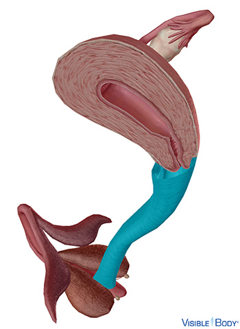
Cervix uteri
The lower part of the uterus constricts into a segment called the cervix, which leads to the vagina. See it in 3D!
System: Reproductive
Region: Pelvis
Function: The cervix is the passageway for menstrual flow, for entering sperm, and for childbirth. Glands in the mucous membrane of the cervix secrete a clear, viscous, alkaline mucus that changes character at different times during a female’s menstrual cycle.
Pathologies: Endometriosis, genital warts, gonorrhea, HPV, infertility, pelvic inflammatory disease, trichomoniasis, uterine fibroids
Vagina
The vagina extends down from the cervix, the lower part of the uterus, to the vestibule, which is part of the vulva and the external genitalia. It sits behind the bladder and in front of the rectum. See it in 3D!
System: Reproductive
Region: Pelvis
Function: The vagina has three core functions: it carries menstrual flow outside the body, it receives the male penis during sexual intercourse, and it serves as a birth canal during labor.
Pathologies: Chlamydia infection, genital herpes, genital warts, HPV, infertility, syphilis, trichomoniasis, yeast infections
Vulva
The external genitalia (vulva) of the female reproductive system include the mons pubis, the labia majora and labia minora, the clitoris and prepuce (clitoral hood), the vestibule of the vagina, urethral orifice, and the greater vestibular glands.
System: Reproductive
Region: Pelvis
Pathologies: Genital warts
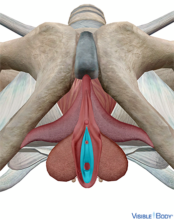
Vestibulum vaginae
The vestibule encompasses the vaginal orifice, the external urethral orifice, and in some females, the hymen. Lateral to the vaginal orifice are masses of erectile tissue known as the bulbs of the vestibule. When a female is sexually aroused, each bulb engorges with blood and swells around the vaginal opening.
System: Reproductive
Region: Pelvis
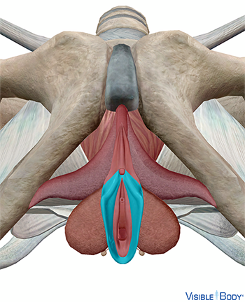
Labia minora pudendi
The labia minora are situated between the labia majora and extend from the clitoris obliquely downward, laterally, and backward for about 4 cm on either side of the orifice of the vagina, enclosing the vestibule. Unlike the labia majora, the labia minora lack pubic hair.
System: Reproductive
Region: Pelvis
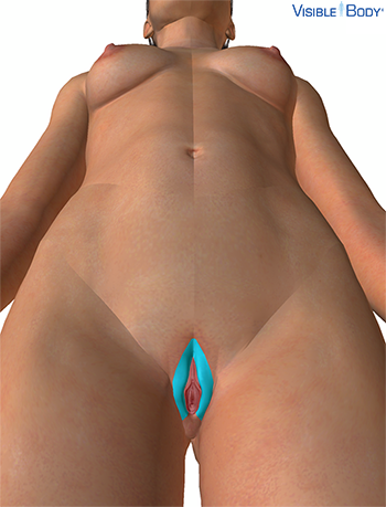
Labia majora
The two labia majora have an outer pigmented surface covered with pubic hair and an inner surface well-endowed with sebaceous (oil) and sudoriferous (sweat) glands.
System: Integumentary, Reproductive
Region: Pelvis
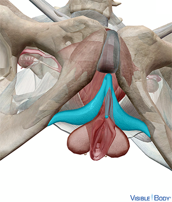
Clitoris
Developmentally homologous to the penis, the clitoris consists of two corpora cavernosa composed of erectile tissue enclosed in a dense layer of fibrous membrane.
System: Reproductive
Region: Pelvis
Function: The clitoris has an extremely high concentration of nerve endings. It can engorge with blood when stimulated and functions in female sexual arousal.
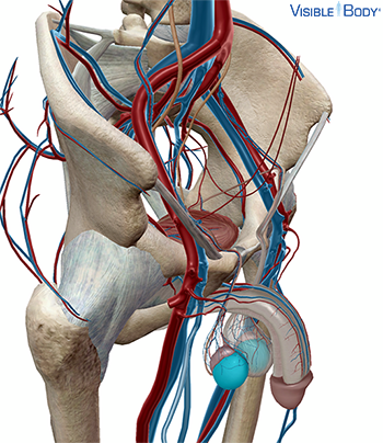
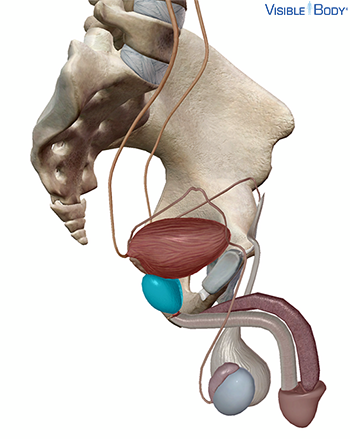
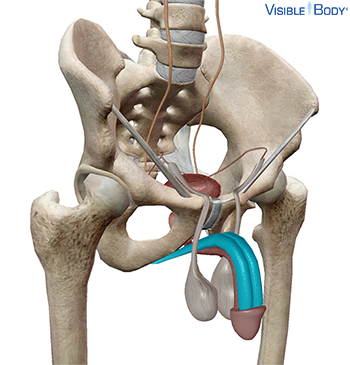
The testes constantly produce sperm—the male sex cells. Sperm are produced in seminiferous tubules inside the testes through a process called spermatogenesis.
System: Reproductive
Region: Pelvis
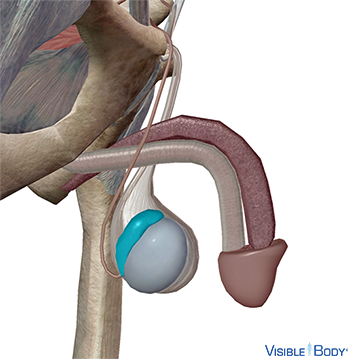
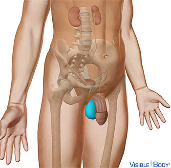
Testis
The testes (or testicles) are the male gonads and sit below the penis within a sac called the scrotum. See it in 3D!
System: Endocrine, Reproductive
Region: Pelvis
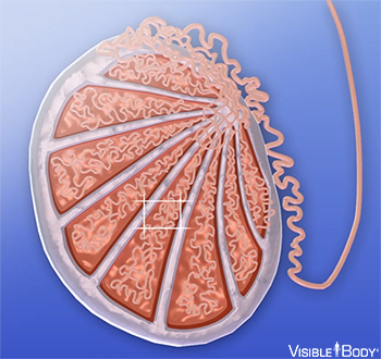
Function: The testes generate sperm, the male sex cells, as well as testosterone and other sex hormones. The production of sperm is constant and occurs within numerous lobules in each testis.
Pathologies: Infertility
Inside testes are coiled tubes called seminiferous tubules. Production begins with the seminiferous tubules where stem cells, called spermatogonia, develop into immature sperm. Each 46-chromosome spermatogonium divides through mitosis to produce primary spermatocytes. These cells divide by meiosis to become 23-chromosome cells called secondary spermatocytes that develop into spermatids.
System: Reproductive
Region: Pelvis
Epididymis
The epididymis is the duct of the male reproductive system that attaches directly to the testis. It is part of the male internal genitalia. The epididymis sits directly on top of each testis.
System: Reproductive
Region: Pelvis
Function: Sperm from the testis mature as they move through the coiled duct of the epididymis. During sexual intercourse and ejaculation, they are expelled into the vas deferens.
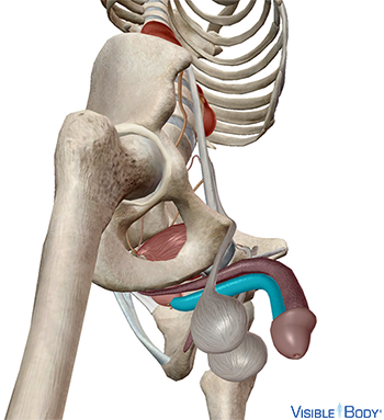
Vas deferens
The vas deferens, also known as the ductus deferens, is one of the ducts of the male reproductive system. The vas deferens serves as the excretory duct of the testis and is the continuation of the epididymis.
System: Reproductive
Region: Pelvis
Function: The vas deferens pushes the sperm up over the bladder and down toward the prostate gland. There, the vas deferens joins the ends of the seminal vesicles (accessory reproductive glands) to form the ejaculatory ducts.
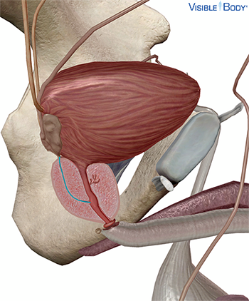
Ductus ejaculatorius
The ducts of the male reproductive system include two ejaculatory ducts, one associated with each testis.
System: Reproductive
Region: Pelvis
Function: The ejaculatory ducts receive seminal fluid from the vesicles, pass through the prostate, and move semen into the urethra.
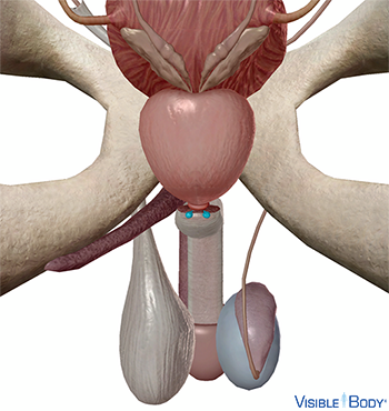
Semen is a mixture of seminal fluid produced by accessory glands and sperm produced by the testes. Sperm cells depend on seminal fluid to keep them moving and alive. This fluid is produced during ejaculation by accessory glands: the seminal vesicles, the prostate, and the bulbourethral glands.
System: Reproductive
Region: Pelvis
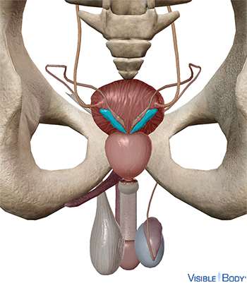
Glandula vesiculosa
The seminal vesicles, two saclike structures, sit close behind the bladder and extend toward the bladder. There they each join one of the vas deferens to form the ejaculatory ducts.
System: Reproductive
Region: Pelvis
Function: The vesicles secrete a whitish-brown fluid containing sugars, prostaglandins, and other substances that makes up two-thirds of the semen volume.
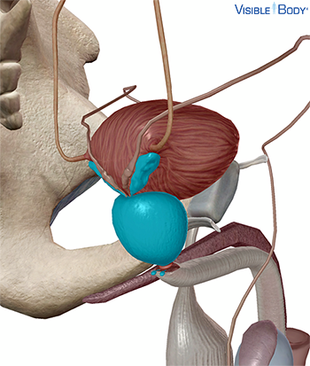
Prostata
The prostate, located under the bladder and above the start of the penis, contains the ejaculatory ducts and the prostatic urethra. See it in 3D!
System: Reproductive
Region: Pelvis
Function: As semen enters the urethra, the prostate secretes enzymes that help activate the sperm.
Pathologies: Enlarged prostate (BPH), prostate cancer
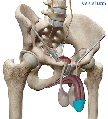
Glandulae bulbourethreales
The bulbourethral glands (or Cowper’s glands) are pea-sized, with single ducts that connect to the urethra where it emerges from the prostate.
System: Reproductive
Region: Pelvis
Function: These glands add mucus that helps with sperm motility.
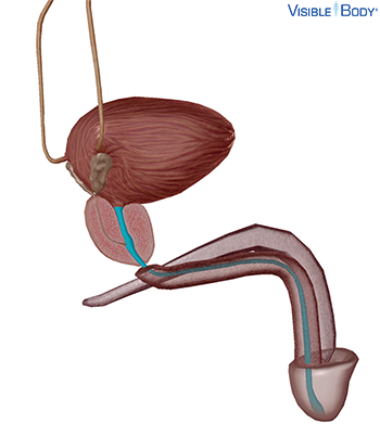
Urethra
The male urethra extends from the bladder, through the prostate, to the external orifice at the end of the penis. It receives additional seminal fluids from the prostate before it expels semen out of the body.
System: Urinary, Reproductive
Region: Pelvis
Pathologies: Urinary tract infections
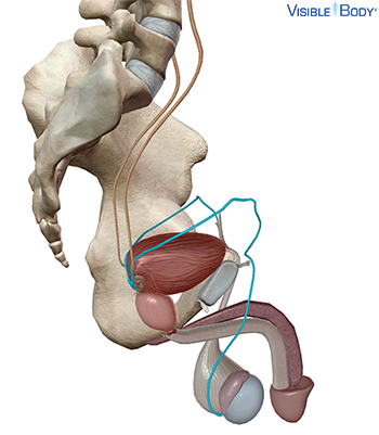
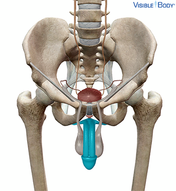
Penis
The penis is part of the male external genitalia, suspended from the body at the front and sides of the pubic arch. Internally, the penis consists of three connected columns of tissue.
System: Reproductive
Region: Pelvis
Function: During sexual arousal, the erectile tissue of the penis fills with blood and the penis stiffens, allowing it to penetrate the vagina during coitus. Ejaculation delivers semen at or near the cervix, the passage to the uterus.
Pathologies: Chlamydia infections, erectile dysfunction, genital herpes, genital warts, gonorrhea, HPV, syphilis, trichomoniasis
Corpus cavernosum
The paired corpora cavernosa extend together from the root of the penis through the body. Together, they form the greater part of the penis.
System: Reproductive
Region: Pelvis
Function: The corpus spongiosum and corpora cavernosa consist of sponge-like erectile tissue containing spaces that can temporarily fill with blood from the deep and dorsal arteries of the penis. These structures engorge with blood and become erect when a male is sexually aroused.
Corpus spongiosum
The corpus spongiosum runs along the underside of the cavernosa. It contains the spongy urethra and expands past the body of the penis to form the glans penis (the tip).
System: Reproductive
Region: Pelvis
Function: The corpus spongiosum and corpora cavernosa consist of sponge-like erectile tissue containing spaces that can temporarily fill with blood from the deep and dorsal arteries of the penis. These structures engorge with blood and become erect when a male is sexually aroused.
Glans penis
The glans penis is the tip of the penis, the external genital organ of the male reproductive system. It arises as the anterior end of the corpus spongiosum and is expanded in the form of a flattened cone. In an uncircumcised male, the loose and retractable skin of the prepuce (foreskin) covers a variable amount of the glans.
System: Reproductive
Region: Pelvis
The testes (or testicles) are the male gonads and sit below the penis within a sac called the scrotum.
System: Integumentary, Reproductive
Region: Pelvis
Pregnancy is a series of events through which a fertilized egg implants, becomes an embryo, and develops into a fetus.
System: Reproduction
Region: Pelvis
Function: The resulting offspring carries genetic information from a male and female into a new generation.
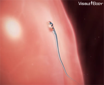
During sexual intercourse, some sperm ejaculated from the male penis swim up through the female vagina and uterus toward an oocyte (egg cell) floating in one of the uterine tubes. The sperm and the egg are gametes. They each contain half the genetic information necessary for reproduction. When a sperm cell penetrates and fertilizes an egg, that genetic information combines.
System: Reproductive
Region: Pelvis
The 23 chromosomes from the sperm pair with 23 chromosomes in the egg, forming a 46-chromosome cell called a zygote. The zygote starts to divide and multiply. As it travels toward the uterus it divides to become a blastocyst, which will burrow into the uterine wall.
System: Reproductive
Region: Pelvis
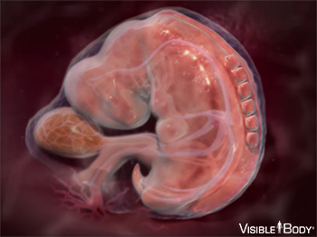
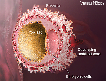
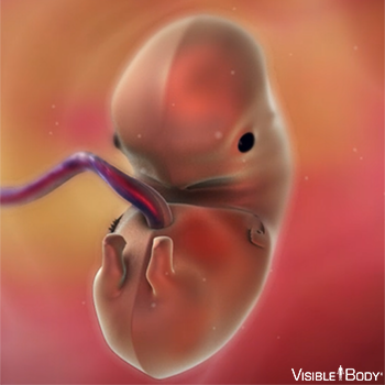
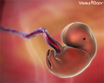
Fifteen days after conception marks the beginning of the embryonic period. The embryo contains a flat embryonic disc that now differentiates into three layers: the endoderm, the mesoderm, and the ectoderm. All organs of the human body derive from these three tissues. They begin to curve and fold and to form an oblong body. By week four, the embryo has a distinct head and tail and a beating heart. Over the next six weeks, limbs, eyes, brain regions, and vertebrae form.
System: Reproductive
Region: Pelvis
At day 15 after conception, the cells that will form the embryo become an embryonic disc. Other cells begin to form support structures. The yolk sac, on one side of the disc, will become part of the digestive tract. On the other side, the amnion fills with fluid and will surround the embryo as it develops.
System: Reproductive
Region: Pelvis
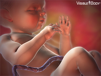
By the end of week 10, the embryo is a fetus. From week 10 of pregnancy, the fetus grows inside the uterus, fueled by nutrient-rich blood supplied by the umbilical cord. Bones, muscles, skin, and connective tissues form. Body systems develop. Limbs and facial features take shape.
System: Reproductive
Region: Pelvis
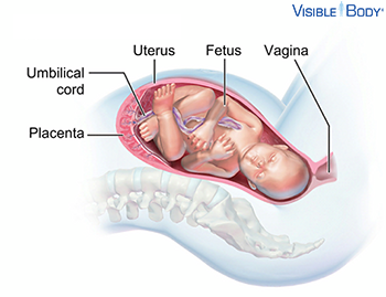
Growth is supplied by nutrients diffusing from the maternal blood into the fetal blood vessels in the placenta. The umbilical cord carries this blood, rich in oxygen and nutrients, to the fetus.
System: Reproductive
Region: Pelvis
The placenta provides oxygen and nutrients to the fetus and removes waste products from the fetus’ blood.
System: Reproductive
Region: Pelvis
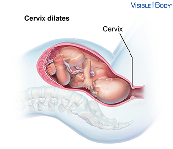
Around week 36 (usually), the process of labor begins. In the first stage, dilation, hormones stimulate downward contractions of the uterine walls. The contractions push the head of the fetus against the cervix at the lower end of the uterus. The cervix dilates.
System: Reproductive
Region: Pelvis
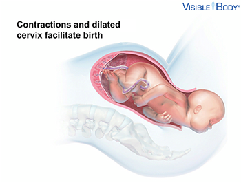
In the second stage, expulsion, powerful contractions push the head and the rest of the body through the dilated cervix, and out through the vagina and the vulva. The baby is born.
System: Reproductive
Region: Pelvis
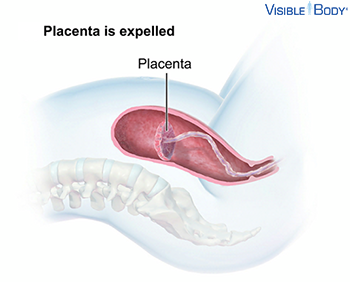
Contractions detach the placenta from the uterus and expel it.
System: Reproductive system
Region: Pelvis