


The lower respiratory system, or lower respiratory tract, consists of the trachea, the bronchi and bronchioles, and the alveoli, which make up the lungs. These structures pull in air from the upper respiratory system, absorb the oxygen, and release carbon dioxide in exchange. Other structures, namely the thoracic cage (or rib cage) and the diaphragm, protect and support these functions.

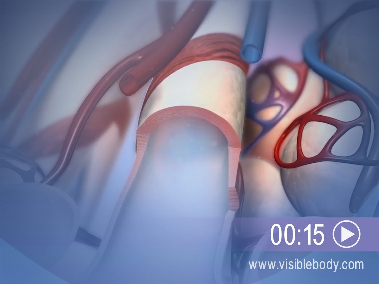
The trachea is a tube less than an inch in diameter, covered by cartilaginous rings. It extends from the bottom of the larynx down behind the sternum, until it branches into smaller tubes, the bronchi. During inhalation, air filtered and warmed by the upper respiratory system passes from the pharynx and larynx into the trachea, then down to the bronchi and into the lungs. Deoxygenated air from the lungs passes back up through the trachea during exhalation. The cartilaginous rings support the tube of the trachea and prevent it from over-expanding or from collapsing, like when you suck on a straw too hard. They are C-shaped, with a gap on the posterior side. This allows the trachea to bend when the esophagus presses against it as food is swallowed.
The tubes of the primary bronchi branch off from the bottom of the trachea. These branches subdivide further into secondary and tertiary bronchi and then into the bronchioles. These progressively smaller airways deliver oxygen-rich air from the trachea to the lungs. During exhalation, deoxygenated air (now rich with carbon dioxide) leaves the lungs by the reverse route. When we exercise, relaxation of smooth muscle in the bronchioles causes them to dilate. This bronchodilation allows greater ventilation. Allergic reactions and histamines cause the opposite effect, bronchoconstriction.
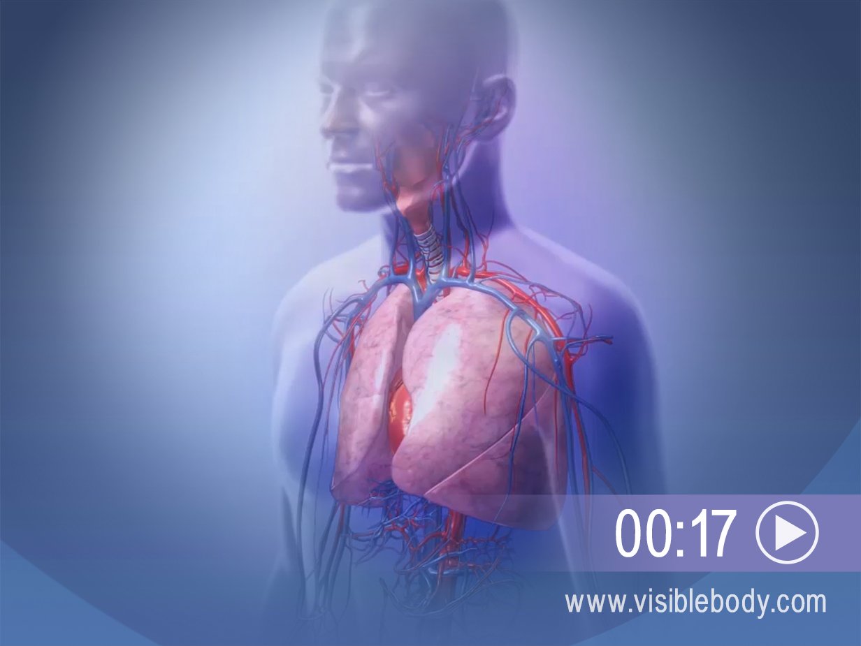
The lungs are responsible for gas exchange between the air we breathe and our bodies. They are protected inside the thoracic cage. The left lung has two lobes and is slightly smaller in volume than the right. It curves in at the cardiac notch to accommodate the heart. The right lung has three lobes. It is slightly shorter, because the diaphragm muscle sits higher below it to accommodate the liver. During inhalation, air flows into the lungs through the bronchi and bronchioles. Oxygen from the air is then absorbed into the bloodstream: it passes through millions of microscopic sacs, the alveoli, into surrounding capillaries. Carbon dioxide waste diffuses the opposite way, from the capillaries to the alveoli. The lungs expel the deoxygenated air during exhalation.
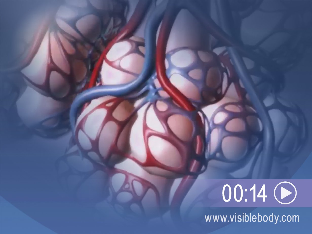
Alveoli are microscopic air sacs served by the bronchioles. Hundreds of millions of alveoli exist inside each lung. They are the terminal ends of the respiratory tract and the sites of external respiration—the exchange of gases between the air and the bloodstream. During inhalation, the alveoli fill with air from the bronchioles.
Oxygen diffuses through the alveoli into networks of pulmonary capillaries that surround them, and is pumped through the bloodstream. Carbon dioxide from deoxygenated blood diffuses from the capillaries into the alveoli, and is expelled through exhalation.

The lungs sit atop the diaphragm, a muscle that forms the floor of the thoracic cavity. The action of the diaphragm is key to the physical process of breathing. During inhalation, the diaphragm contracts and moves inferiorly, toward the abdominal cavity. This allows the volume of the thoracic cavity and the lungs to increase. It also explains why your abdomen puffs out when you take a deep breath. During normal exhalation, the diaphragm relaxes (along with the external intercostal muscles). The thoracic cavity and lungs decrease, and air is expelled.
