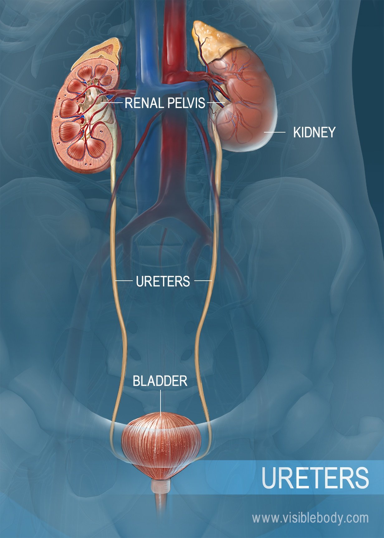
The kidneys, ureters, bladder, and urethra are the primary structures of the urinary system. They filter blood and remove waste from the body in the form of urine. The size and position of lower urinary structures vary with male and female anatomy.

The kidneys are bean-shaped organs situated on the back of the abdominal wall, behind the peritoneum. The right kidney sits slightly lower than the left to accommodate the liver. The kidneys filter blood (supplied by the renal arteries) to remove unwanted substances. They also secrete waste into the urine.

Urine drains from the renal pelvis of each kidney into the ureters. The ureters are long, thin tubes made of smooth muscle. Contractions of the smooth muscle push urine down through the ureters and into the bladder. In adults, the ureters are 25–30 cm long, about the length of a 12-inch ruler.

Urine flows through the ureters into the urinary bladder. In women, the bladder is located in front of the vagina and below the uterus. In men, the bladder sits in front of the rectum and above the prostate gland. The wall of the bladder contains folds called rugae, and a layer of smooth muscle called the detrusor muscle. As urine fills the bladder, the rugae smooth out to accommodate the volume. The detrusor relaxes to hold the urine, then contracts for urination. An adult bladder is full at about half a liter, or about two cups.

Urine produced in the kidneys passes through the ureters, collects in the bladder, and is then excreted through the urethra. In females, the urethra is narrow and about 4 cm long, significantly shorter than in males. It extends from the bladder neck to the external urethral orifice in the vestibule of the vagina.

In males, the urethra is about 17.5–20 cm, four or five times as long as in females. The male urethra is divided into three sections: the prostatic urethra (the widest portion), the membranous urethra (the narrowest portion), and the spongy urethra (the longest portion). It extends from the bladder neck through the prostate and the penis to the external urethral orifice. In men, both urine and semen pass out of the body through the urethra.
A description of the organs of the urinary system from the 1918 edition of Gray's Anatomy of the Human Body.
An overview of the urinary system from the Cleveland Clinic.
Visible Body Web Suite provides in-depth coverage of each body system in a guided, visually stunning presentation.
When you select "Subscribe" you will start receiving our email newsletter. Use the links at the bottom of any email to manage the type of emails you receive or to unsubscribe. See our privacy policy for additional details.