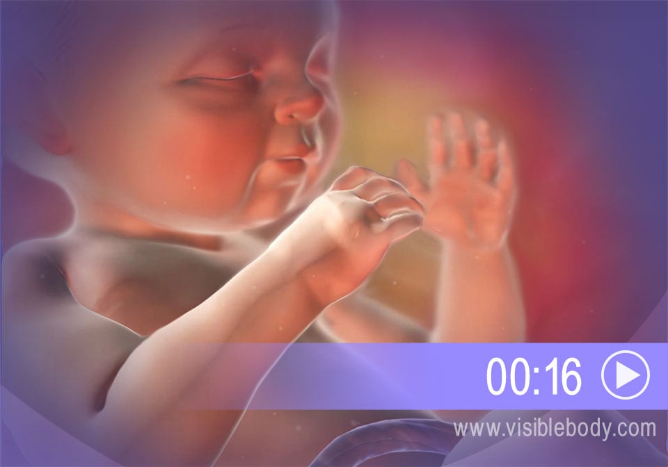
In the reproductive process, a male sperm and a female egg provide the information required to produce another human being. Conception occurs when these cells join as the egg is fertilized. Pregnancy begins once the fertilized egg implants in the uterus. The embryo grows and becomes surrounded by structures that provide support and nourishment. Eyes, limbs, and organs appear as the embryo develops into a fetus. The fetus grows inside the uterus until pregnancy ends with labor and birth. By then all body systems are in place—including the reproductive system that can one day help produce another human being.
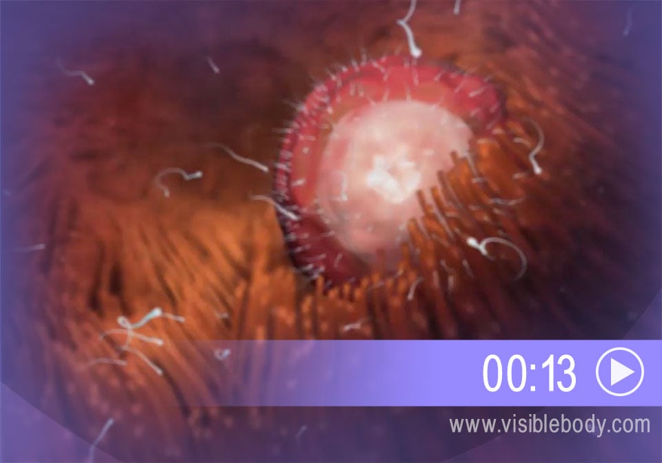
During sexual intercourse, some sperm ejaculated from the male penis swim up through the female vagina and uterus toward an oocyte (egg cell) floating in one of the uterine tubes. The sperm and the egg are gametes. They each contain half the genetic information necessary for reproduction. When a sperm cell penetrates and fertilizes an egg, that genetic information combines. The 23 chromosomes from the sperm pair with 23 chromosomes in the egg, forming a 46-chromosome cell called a zygote. The zygote starts to divide and multiply. As it travels toward the uterus it divides to become a blastocyst, which will burrow into the uterine wall.
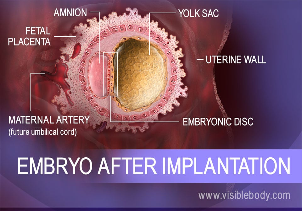
A fertilized egg, or zygote, takes about five days to reach the uterus from the uterine tube. As it moves, the zygote divides and develops into a blastocyst, with an inner mass of cells and a protective outer ring. The blastocyst attaches to the wall of the uterus and gradually implants itself into the uterine lining. During implantation, its cells differentiate further. At day 15 after conception, the cells that will form the embryo become an embryonic disc. Other cells begin to form support structures. The yolk sac, on one side of the disc, will become part of the digestive tract. On the other side, the amnion fills with fluid and will surround the embryo as it develops. Other cell groups initiate the placenta and umbilical cord, which will bring in nutrients and eliminate waste.
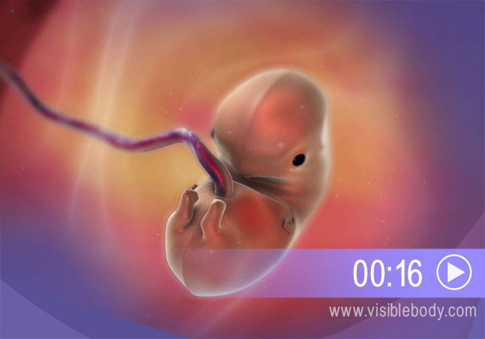
Fifteen days after conception marks the beginning of the embryonic period. The embryo contains a flat embryonic disc that now differentiates into three layers: the endoderm, the mesoderm, and the ectoderm. All organs of the human body derive from these three tissues. They begin to curve and fold and to form an oblong body. By week 4, the embryo has a distinct head and tail and a beating heart. Over the next six weeks, limbs, eyes, brain regions, and vertebrae form. Primitive versions of all body systems appear. By the end of week 10, the embryo is a fetus. (Note: Pregnancy is often measured in terms of gestational age—age of the fetus starting with the first day of a woman’s last menstrual period—and embryonic or fetal age—actual age of the growing fetus. We are referring to the gestational age of the fetus.)
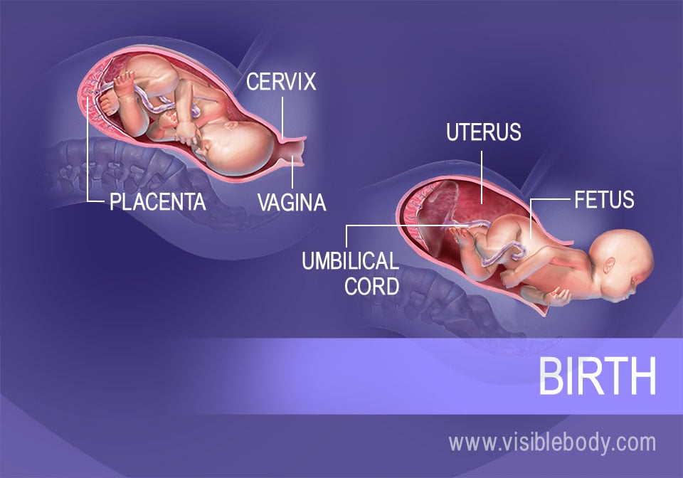
From week 10 of pregnancy, the fetus grows inside the uterus, fueled by nutrient-rich blood supplied by the umbilical cord. The placenta provides oxygen and nutrients to the fetus and removes waste products from the fetus’ blood. Bones, muscles, skin, and connective tissues form. Body systems develop. Limbs and facial features take shape. Around week 36 (usually), the process of labor begins. In the first stage, dilation, hormones stimulate downward contractions of the uterine walls. The contractions push the head of the fetus against the cervix at the lower end of the uterus. The cervix dilates. In the second stage, expulsion, powerful contractions push the head and the rest of the body through the dilated cervix, and out through the vagina and the vulva. The baby is born. Further contractions expel the placenta to complete the placental stage.
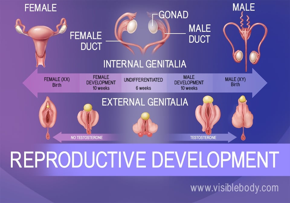
Reproductive structures begin to form in the embryonic stage. By week 6, gonads and genitalia are present but undifferentiated. Whether they become male or female is determined by one chromosome delivered by the sperm. This pair contains an X sex chromosome from the female egg and either an X or a Y sex chromosome from the male sperm. If the chromosome pair is XY, the gonads develop into testes starting in week 7. If the chromosome pair is XX, the gonads become ovaries starting in week 8. Testes secrete testosterone, forming male genitalia around week 10. Without testosterone, female genitalia form. All reproductive structures are in place at birth or shortly after. At puberty, an increase in sex hormones will grow them to their adult size and reproductive capability.
A description of the embryo at different stages of its growth from the 1918 edition of Gray’s Anatomy of the Human Body.
An article in Science Daily on a research study that involves creating a synthetic model of the placenta to better understand pregnancy.
Visible Body Web Suite provides in-depth coverage of each body system in a guided, visually stunning presentation.
When you select "Subscribe" you will start receiving our email newsletter. Use the links at the bottom of any email to manage the type of emails you receive or to unsubscribe. See our privacy policy for additional details.