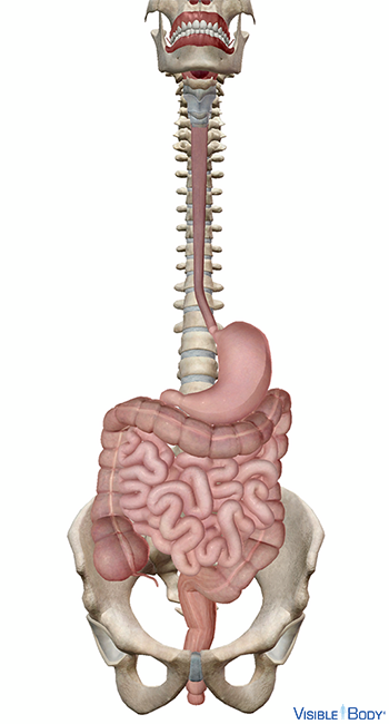
The alimentary canal is a musculomembranous tube that extends from the mouth to the anus.
System: Digestive
Region: Head, Neck, Thorax, Abdomen, Pelvis
Function: It is through mechanical and chemical processes that occur in this passageway that consumed food is digested, nutrients are extracted, and waste is expelled.
Cavitas oris
The oral cavity (mouth) is an oval-shaped cavity located anterior to the pharynx at the beginning of the alimentary canal, where the process of digestion is initiated. See it in 3D!
System: Digestive
Region: Head
Function: Food is masticated in the oral cavity by the action of the teeth and tongue and mixed with salivary gland secretions, which contain enzymes that begin to break down carbohydrates.
Pathologies: Canker sores, cold sores, gum disease
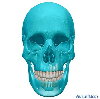
Dentes
The permanent teeth are 32 bone-like structures embedded in the maxillae and the mandible of the skull.
System: Skeletal
Region: Head
Function: Teeth are used for mastication.
Pathologies: Tooth decay
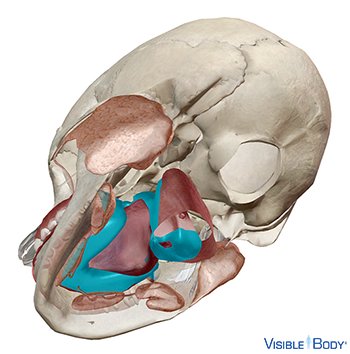
Glandulae salivariae
Salivary glands (r, l) are accessory exocrine glands of the digestive system. They are situated in the oral cavity underneath the mucosa.
System: Digestive
Region: Head
Function: Three pairs of extrinsic salivary glands aid in digestion: parotid, sublingual, and submandibular (also called submaxillary). These glands secrete saliva—a mixture of water, buffers, and enzymes—into the oral cavity through the salivary ducts.
Pathologies: Rheumatoid arthritis
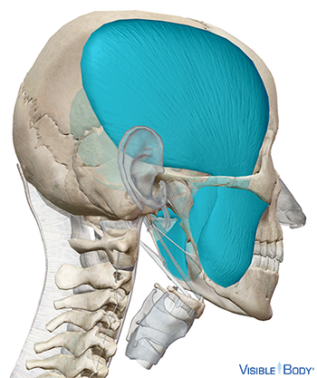
The muscles of mastication are a subgroup of the muscles of the head. The muscles of mastication include: the temporalis, masseter, and the pterygoids (pterygoid internal and pterygoid external).
System: Muscular
Region: Head
Function: These muscles work together to act on the temporomandibular joint and produce the motions involved in mastication.
Pathologies: Muscular dystrophy, myositis
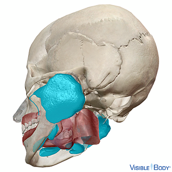
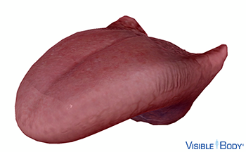
Lingua
The tongue is made up of four intrinsic muscles: the superior lingualis, the inferior lingualis, the vertical lingualis, and the transverse lingualis. These muscles work together to give the tongue its great flexibility. See it in 3D!
System: Digestive, Muscular
Region: Head
Function: The tongue manipulates food in the oral cavity and contains taste buds, the sensory receptors for taste.
Pathologies: Muscular dystrophy, myositis
Pharynx
The pharynx is a 12.5-cm conical musculomembranous tube. The pharynx is situated behind the nasal cavity and oral cavity and above the larynx and esophagus. The pharynx is divided into three segments: the nasopharynx, the oropharynx, and the laryngopharynx. See it in 3D!
System: Digestive
Region: Neck
Function: The pharynx functions as part of the alimentary canal and as an airway in the upper respiratory system.
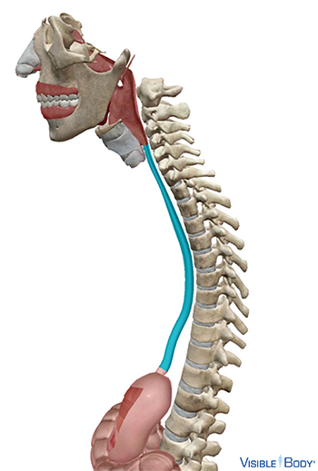
Esophagus
The esophagus is an approximately 23-to-25-cm-long hollow muscular tube extending from the pharynx to the stomach; it is the narrowest part of the alimentary canal. See it in 3D!
System: Digestive
Region: Thorax
Function: Peristaltic waves in the esophagus force the food mass (bolus) toward the stomach as part of the process of swallowing.
Pathologies: GERD, heartburn, hiatal hernia
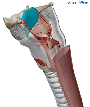
Epiglottis
The epiglottis is one of the nine cartilages that joint to form the laryngeal skeleton (also known as the larynx or voice box), which is attached to structures of the axial skeleton. See it in 3D!
System: Skeletal
Region: Neck
Function: The epiglottis is usually directed upward toward the pharynx, but during swallowing, muscles pull it down to close the entry to the larynx and prevent food from entering the trachea.
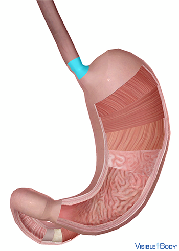
Sphincter cardiacus
The cardiac or gastroesophageal sphincter regulates the passage of food from the esophagus into the stomach.
System: Digestive
Region: Abdomen
Function: When the peristaltic wave passing through the esophagus reaches the gastroesophageal opening, the cardiac sphincter opens, allowing the bolus to pass into the stomach.
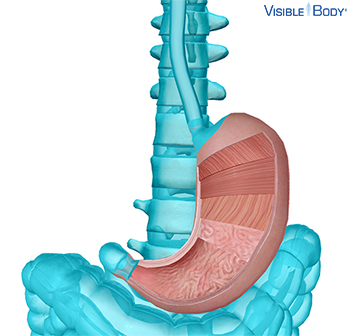
Gaster
The stomach is the most dilated part of the alimentary canal; situated between the esophagus and the beginning of the small intestine, it is a principal organ of the digestive system. See it in 3D!
System: Digestive
Region: Abdomen
Function: Along with providing food storage, the stomach breaks down ingested food into chyme, both mechanically and by the action of stomach muscles and chemically through secreted acids and enzymes that constitute the gastric juice emitted by the stomach’s mucosa.
Pathologies: GERD, hiatal hernia, peptic ulcer
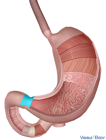
M. sphincter pylori
The pyloric sphincter is a valve that regulates the passage of chyme from the pylorus of the stomach to the duodenum.
System: Digestive
Region: Abdomen
Function: A peristaltic wave triggers the pyloric sphincter to release a few milliliters of chyme at a time into the duodenum.
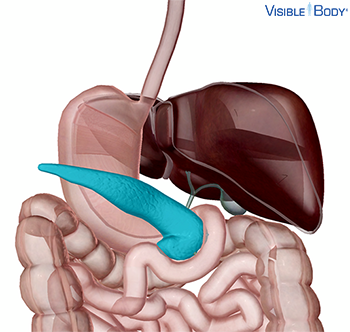
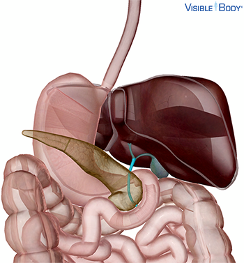
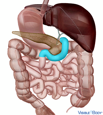
Pancreas
The pancreas is primarily an exocrine gland that secretes pancreatic juice, an important digestive fluid. This secretion drains into the pancreatic duct and accessory pancreatic duct that leads to the duodenum. See it in 3D!
System: Digestive
Region: Abdomen
Function: The mixture of digestive enzymes, water, and electrolytes called pancreatic juice, produced in acinar and epithelial cells, drains into the pancreatic duct which extends transversely from the tail toward the head.
Pathologies: Pancreatitis, prediabetes
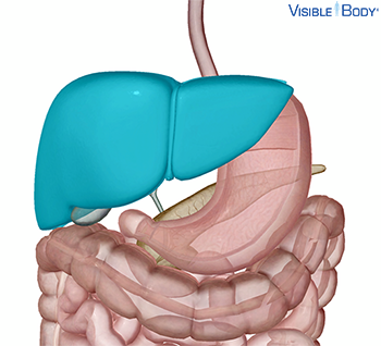
Hepar
The 1.2- to 1.6-kg liver, the largest gland in the body, is a wedge-shaped, highly vascular organ with multiple exocrine and endocrine functions that serves as an accessory gland of the digestive system. See it in 3D!
System: Digestive
Region: Abdomen
Function: The liver’s exocrine secretion (bile) facilitates the digestion of fats and the removal of excess cholesterol from the blood.
Pathologies: Cirrhosis, hepatitis A, hepatitis B, hepatitis C, jaundice
Ductus choledochus
The common bile duct is part of the ductal system that allows bile to flow from the liver and gall bladder into the duodenum to aid in digestion.
System: Digestive
Region: Abdomen
Function: When the sphincter of Oddi is closed, bile flows up the common bile duct and through the cystic duct for storage in the gall bladder.
Pathologies: Gallstones
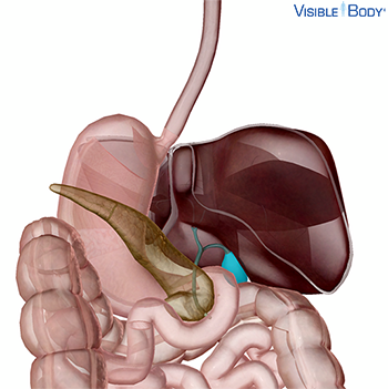
Vesica biliaris
The gall bladder is a conical or pear-shaped musculomembranous sac 7 to 10 cm in length that serves as a reservoir for bile secreted by the liver. See it in 3D!
System: Digestive
Region: Abdomen
Function: Bile generated by the liver moves out of the common hepatic duct through the common bile duct into the duodenum; when the sphincter of Oddi is closed, the bile is stored and concentrated in the expandable gall bladder.
Pathologies: Gallstones
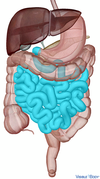
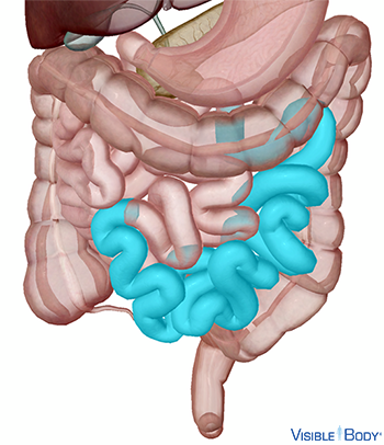
Intestinum tenue
The small intestine is an approximately 7-m-long convoluted portion of the alimentary canal within the abdominal cavity. See it in 3D!
System: Digestive
Region: Abdomen
Function: It has a vital role in chemical digestion and the absorption of water, nutrients, and ions.
Pathologies: Adhesions, Clostridium difficile infections, Crohn's Disease, diarrhea, gastroenteritis, hernia, intestinal obstruction, lactose intolerance, peptic ulcer
Peristaltic waves push food down from the esophagus to the stomach, where it is churned and passed into the small intestine. Rhythmic smooth muscle contraction pushes chyme through the small and large intestines.
System: Digestive
Region: Thorax, Abommen, Pelvis
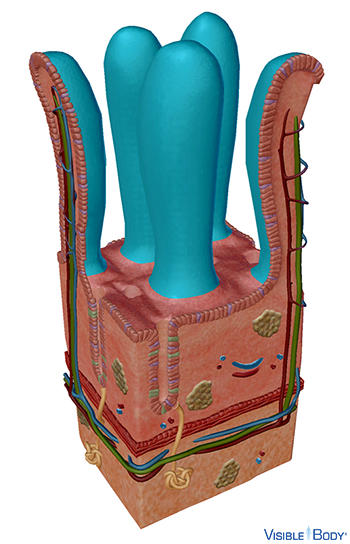
Villi intestinales
The surface of the small intestine is composed of villi, small fingerlike projections that extend into the lumen and increase the intestinal wall’s surface area, allowing for increased nutrient absorption.
System: Digestive
Region: Abdomen
Function: As chyme passes through the lumen, the villi absorb nutrients through columnar epithelial cells of the mucosa, called absorptive cells or enterocytes.
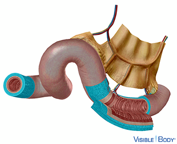
Duodenum
The duodenum is a short, wide stretch of the small intestine that receives chyme from the stomach and digestive enzymes from the liver and the pancreas.
System: Digestive
Region: Abdomen
Function: Chyme passes from the stomach into the duodenum through the pyloric sphincter.
When the sphincter of Oddi is relaxed, bile secreted from the liver and pancreatic juices secreted from the pancreas enter the duodenum to aid in chemical digestion and to reduce acidity.
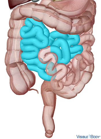
Intestinum jejunum
The jejunum is the middle portion of the small intestine.
System: Digestive
Region: Abdomen
Function: The jejunum is thicker, more vascular, and of a deeper color than the ileum, with large circular folds of submucosa called plicae circulares and villi larger than in the ileum.
Intestinum ileum
The ileum is the longest segment of the small intestine.
System: Digestive
Region: Abdomen
Function: The walls are thinner and less vascular than those of the jejunum, with few plicae circulares that are small and disappear gradually.
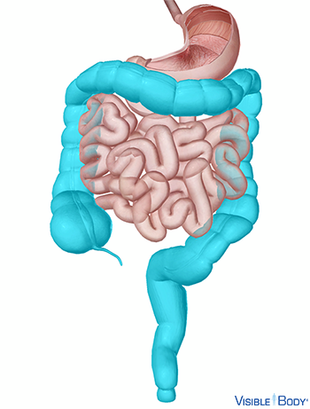
Intestinum crassum
The large intestine is a 1.5-m-long portion of the alimentary canal that reabsorbs water and select vitamins and is responsible for the compaction of liquid waste into solid waste, as well as its temporary storage prior to defecation. See it in 3D!
System: Digestive
Region: Abdomen
Function: The large intestine receives food and some water, vitamins, and minerals from the small intestine. Any of this liquid that it does not absorb it eliminates as solid waste. Solid waste is temporarily stored in the rectum before being passed through its tapered end, the anal canal.
Pathologies: Adhesions, appendicitis, Clostridium difficile infections, colonic polyps, diarrhea, diverticulosis and diverticulitis, hernia, intestinal obstruction, irritable bowel syndrome, ulcerative colitis
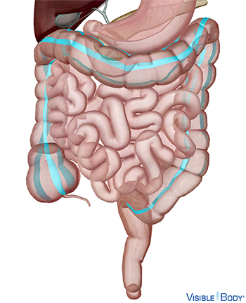
Taeniae coli
Taenia coli are bands of smooth muscle that extend along the surface of the large intestine, aiding in the movement of fecal material through the colon.
System: Digestive
Region: Abdomen, Pelvis
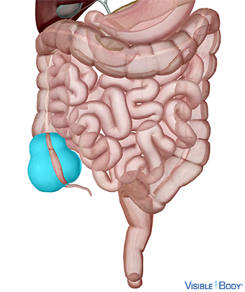
Intestinum caecum
The cecum is a large pouch that is the beginning of the large intestine, the portion of the alimentary canal responsible for final absorption of nutrients and the compaction of liquid waste into solid waste. See it in 3D!
System: Digestive
Region: Abdomen
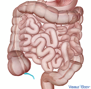
Appendix vermiformis
The appendix is a long, slender tube attached at the base of the cecum with great variability. It varies in length from 2 to 20 cm, with an average of approximately 8 cm. See it in 3D!
System: Digestive
Region: Abdomen
Function: Once considered to be solely a vestigial structure, the appendix is now known to function as part of the lymphatic system, as it contains a large amount of lymphoid aggregates that are part of the widely distributed gut-associated lymphoid tissue (GALT)
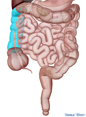
Colon ascendens
The ascending colon is a vertical segment of the colon that passes upward from the cecum to below the right liver lobe.
System: Digestive
Region: Abdomen
Function: As a region of the colon and part of the large intestine, it is responsible for water absorption and the transformation of intestinal materials into feces.
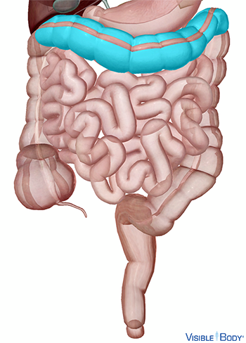
Colon transversum
The transverse colon is a horizontal segment of the colon. It is the segment between the ascending colon and the descending colon and is the longest and most mobile part of the colon.
System: Digestive
Region: Abdomen
Function: As a region of the colon and part of the large intestine, it is responsible for water absorption and the transformation of intestinal materials into feces.
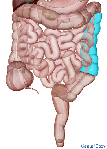
Colon descendens
The descending colon is a vertical segment of the colon that passes downward through the left hypochondriac and lumbar regions.
System: Digestive
Region: Abdomen
Function: It is part of the large intestine, the organ responsible for water absorption and the compaction and storage of solid waste.
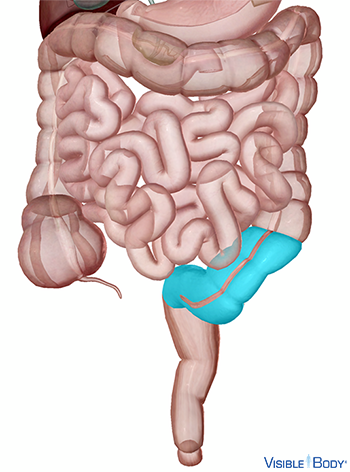
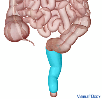
Colon sigmoideum
The sigmoid colon is the shortest and narrowest part of the colon.
System: Digestive
Region: Pelvis
Function: It is located within the region of the pelvis and is responsible for the production and storage of fecal material.
Rectum
The rectum is the final segment of the large intestine and serves primarily to store and expel solid waste.
System: Digestive
Region: Pelvis
Function: When the lower part of the rectum is contracted, its mucous membrane forms longitudinal folds that prevent solid waste from pushing toward the anal canal, thus causing a sensation that elicits the urge to defecate.
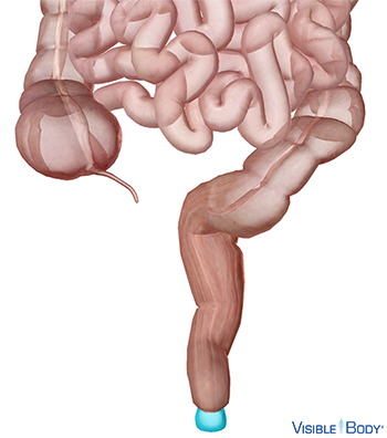
Pars analis recti
The anal canal is the inferior portion of the rectum of the large intestine and the termination of the alimentary canal.
System: Digestive
Region: Pelvis
Function: Its primary function is to temporarily store solid waste that is ready to be eliminated from the body.
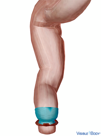
Musculus sphincter ani internus
The internal anal sphincter, one of the pelvic floor muscles, is a muscular ring that surrounds about 2.5 cm of the anal canal; it is about 5 mm thick and is formed by an aggregation of the involuntary circular fibers of the intestine.
System: Muscular
Region: Pelvis
Function: The body expels waste products from digestion through the rectum and anus. This process, called defecation, involves contraction of rectal muscles, relaxation of the internal anal sphincter, and an initial contraction of the skeletal muscle of the external anal sphincter.
Pathologies: Muscular dystrophy, myositis
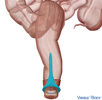
The external anal sphincter, one of the pelvic floor muscles, is a circular sphincter muscle in the anala region of the perineum.
System: Muscular
Region: Pelvis
Function: The body expels waste products from digestion through the rectum and anus. This process, called defecation, involves contraction of rectal muscles, relaxation of the internal anal sphincter, and an initial contraction of the skeletal muscle of the external anal sphincter.
Pathologies: Muscular dystrophy, myositis