
3D Skeletal System: The Shoulder Girdle
Think of how many things you do during the day; you stretch first thing in the morning, lift your coffee cup, wave at people, type, write, text, play Pokémon Go—no matter what it is you do, chances are you're using your arms to do them. And you know what's incredible about that? You wouldn't be able to use your arms without the shoulder girdle!
I'm fascinated by the shoulder girdle, to be honest. It's such an interesting group of structures and presents an even more interesting joint. Let's take a quick look, shall we?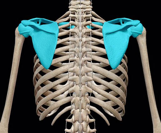
Bones of the Shoulder Girdle
The shoulder girdle is made up of two posterior scapulae and two anterior clavicles. These bones make an incomplete ring around the upper thoracic cage. The medial end of each clavicle articulates with the manubrium and scapulae; each scapula connects to the thoracic cage by muscle only. How crazy is that? The only thing that connects the upper limbs and the scapula to the axial skeleton are the clavicles. That's a pretty big job, wouldn't you say?The scapulae are flat, triangular bones that serve as the attachment sites of many muscles, including the deltoids. Muscles of the back and thorax connect the scapulae to the thoracic cage.
Here's a quick run-down of the girdle's articulations:
|
|
Articulations |
|
Scapula |
Humerus, clavicle |
|
Clavicle |
Scapula, manubrium, cartilage of the first rib |
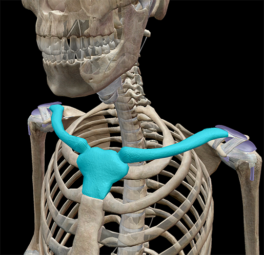 Image from Visible Body Suite.
The Glenoid Cavity and the Shoulder Joint
Hold one of your arms out and rotate it. Your arms have more motility than your legs, despite the fact that the shoulder and hip joints are the same type of joint!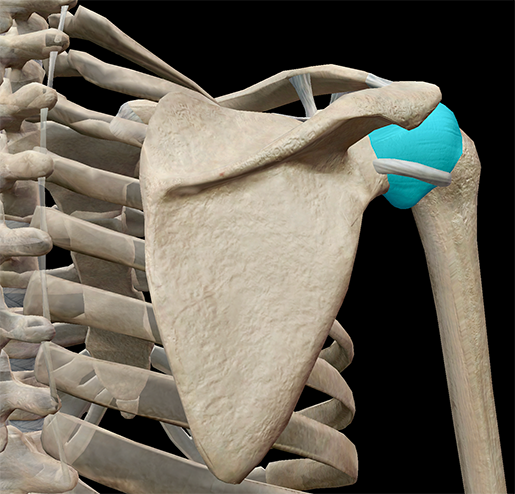
Image from Visible Body Suite.
| Did you know? Ball-and-socket joints are one of the six types of synovial joints in the body. Check out the Learn Site to learn about gliding, hinge, pivot, condyloid, and saddle joints! |
Watch the range of motion of a ball-and-socket joint here:
Landmarks of the Scapula
The scapula is such an interesting bone. I mean, look at it! It's such a cool shape that it's also known as the shoulder blade. You are walking around with two knives permanently strapped to your back. How awesome is that?Landmarks, or characteristics of the bone, have different functions: muscle attachment sites, bone and ligament articulation sites, passage for nerves and vessels, and more. In the image, all bone landmarks are highlighted in different colors.
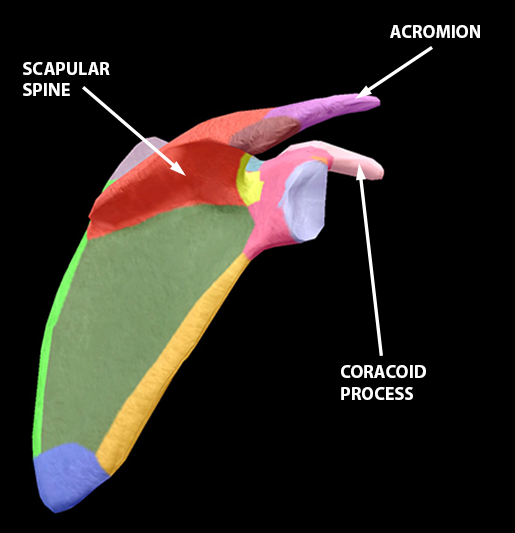
The scapular spine (in pale orange-red) is a prominent plate of bone that serves as the attachment site for portions of the trapezius and the deltoid. At the distal end of the spine is the acromion.
Do you have Visible Body Suite and want to keep the learning going? Study the appendicular skeleton with our premade Flashcard deck! Here's how to open it in VB Suite:
1. Copy this link:
https://apps.visiblebody.com/share/?p=vbhaa&t=4_55710_637636852041160000_162926
2. Use the Share Link button in VB Suite.
3. Paste the link to view the deck.
4. Save the deck to your User Account.
For more premade Flashcard decks on popular topics, check out our whole library here.
Be sure to subscribe to the Visible Body Blog for more anatomy awesomeness!
Are you an instructor? We have award-winning 3D products and resources for your anatomy and physiology course! Learn more here.
Related Posts:
- 3D Skeletal System: Atlas, Axis, and the Atlanto-Axial Relationship
- 3D Skeletal System: Function of the Sphenoid
- 3D Skeletal System: The Pelvic Girdle
Additional Sources:
- Gray's Anatomy (The Acromioclavicular Articulation)

