
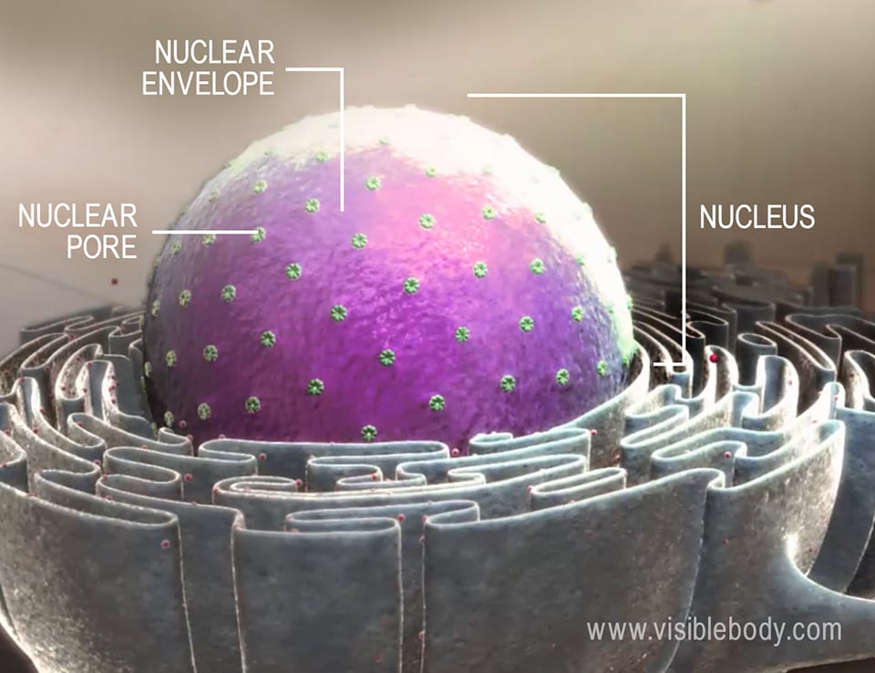
The principal feature that distinguishes a eukaryotic cell from a prokaryotic cell is the presence of a membrane-bound nucleus. This nucleus is the “control center” of the cell that stores all the cell’s genetic information, or DNA. The nuclear membrane, or nuclear envelope, contains channels called pores that regulate the movement of molecules in and out of the nucleus.
The DNA inside the nucleus is organized into chromosomes. At the most basic level, a chromosome is a molecule of DNA that is tightly coiled around proteins called histones. Eukaryotic cells have multiple chromosomes that are linear in shape.
Individual DNA molecules are extremely long, consisting of millions of base pairs (matched nucleotides) each. How do cells store such large and potentially unwieldy molecules? Much like you’d store yarn or thread—wound onto a spool—DNA is compacted, coiled, and folded into a compact shape.

Chromatin consists of all the DNA in the nucleus, as well as its associated proteins. There are three basic layers of chromatin scaffolding that results in a condensed DNA molecule.
The double helix shaped DNA molecule that makes up each chromosome is first coiled around clusters of histone proteins. A unit of around 200 DNA base pairs wound around eight histone proteins makes up the smallest unit of DNA-packing structure, a nucleosome.
The nucleosomes and the linker DNA that connects them, like beads on a string, loop to form more tightly-packed 30-nm solenoid fibers. Then, the 30-nm fibers are coiled further and folded into loops that are tightly packed together. This last stage of scaffolding produces the chromosomes observable in metaphase of mitosis or meiosis.
The process that creates the 30nm fibers is called supercoiling. Supercoiling uses the application of tension to twist a DNA molecule, so it wraps around itself, creating loops.
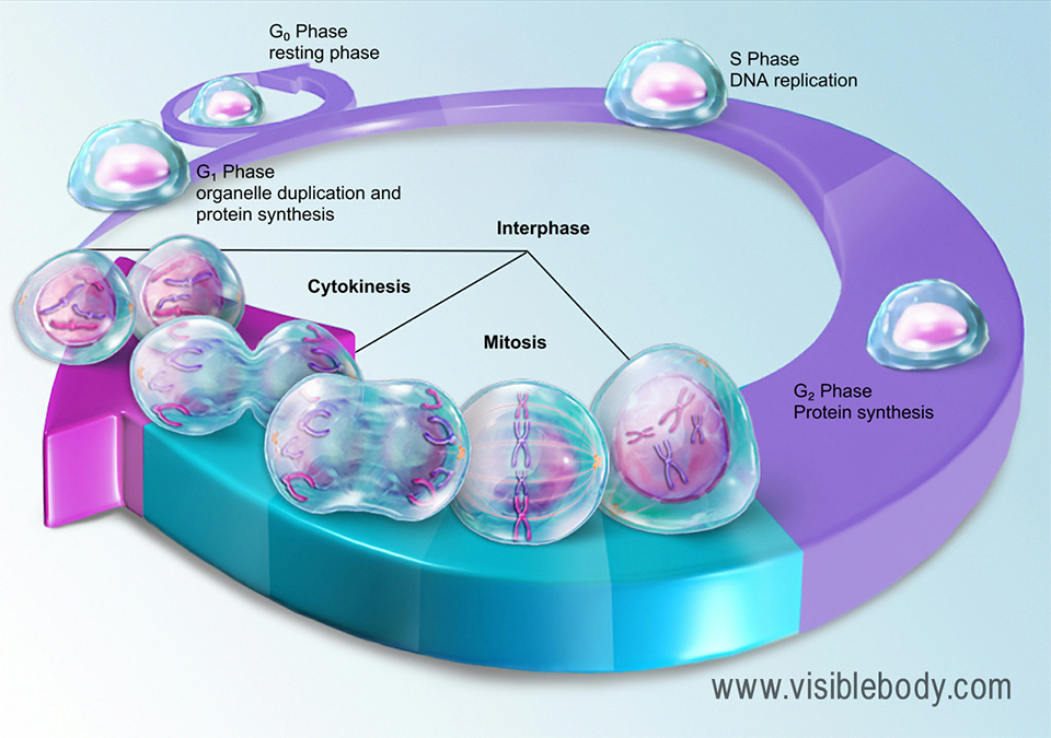
Even with a microscope, individual chromosomes are clearly visible in the nucleus only during the process of cell division (either mitosis or meiosis). This is because the chromatin that makes up the chromosomes is hundreds, or even thousands, of times less condensed during interphase than it is when the cell is actively dividing.
Before mitosis begins, a cell’s DNA replicates itself, making an identical copy of each chromosome.
It is during mitosis or meiosis that the X-shaped structures we usually picture when thinking of chromosomes can be observed.
Each of these X-shaped chromosomes consists of two identical sister chromatids. Basically, you can think of a chromatid as one copy of a chromosome.
The sister chromatids are connected to each other by a region of the chromosome called the centromere. During mitosis, spindle fibers attach to this region, and they eventually pull the sister chromatids apart to form two separate chromosomes, one for each daughter cell.
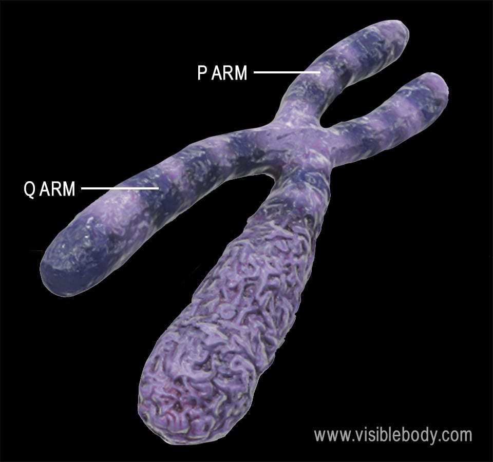
The centromere also divides each chromatid into two regions: the p arm and the q arm. The p arm is the shorter arm, and the q arm is the longer arm. When geneticists describe the location of a gene on a chromosome, they will specify the number of the chromosome (1-22, X, and Y for humans), the arm (p or q), and the gene’s location on the arm.
The somatic cells of the human body—that is, the cells that aren’t gametes, or sex cells—each have 46 chromosomes. They are diploid cells, which means that those 46 chromosomes are organized into 23 pairs. Diploid is sometimes abbreviated as 2n (where n is the number of different chromosomes).
In humans, 22 out of the 23 chromosome pairs are autosomes, or non-sex chromosomes. These are referred to as homologous pairs because the two chromosomes within the pair are the same size and shape and contain the same genes (with potentially different alleles, which are alternate versions of a gene). The 23rd pair is made up of the sex chromosomes. Typically, people who are biologically male have an XY genotype (an X chromosome and a Y chromosome), and people who are biologically female have an XX genotype (two X chromosomes). The XX pair, but not the XY pair, is considered to be a homologous pair.
The sex cells, or gametes, of sexually-reproducing eukaryotic organisms are haploid (1n), meaning they only have 23 unpaired chromosomes. When a sperm cell fertilizes an egg, the resulting zygote is diploid; the combination of the two haploid sex cells is what results in the zygote having the full 46 chromosomes. One chromosome in each pair comes from the mother’s egg cell, and the other comes from the father’s sperm cell.
Telomeres are special regions, located on each end of the chromosome, that consist of repetitive sequences of base pairs. They are typically described as being like the plastic caps on the ends of shoelaces. This is because they protect the ends of chromosomes and keep them from sticking to other chromosomes.
However, the most important function of telomeres is that they allow cells to divide without losing their genetic material. If there were no telomeres, some base pairs would be lost every time DNA replication takes place. Fortunately, it is the telomeres, and not the genes, that get shorter with every cell division. For example, the telomeres in human cells generally lose between 30 and 200 base pairs every time a cell divides.
Normally, a cell in the human body can divide around 50-70 times before it becomes senescent (inactive, no longer dividing) or dies. As humans age, the telomeres on the ends of the chromosomes gradually become shorter.
A special enzyme called telomerase adds telomeres to the ends of replicated chromosomes, meaning that cells with an increased level of this enzyme—like sperm and egg cells, or certain cancer cells—can divide more frequently.
Here’s how the characteristics of eukaryotic chromosomes compare to those of prokaryotic chromosomes:
| Eukaryotic Chromosome | Prokaryotic Chromosome | |
|---|---|---|
| Shape | Linear | Circular |
| Size | Large | Small |
| Number | Multiple | Single |
| Location | Nucleus | Nucleoid (region in cytoplasm) |
| Storage proteins | Histones | Nucleoid-associated proteins |
