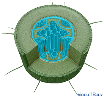
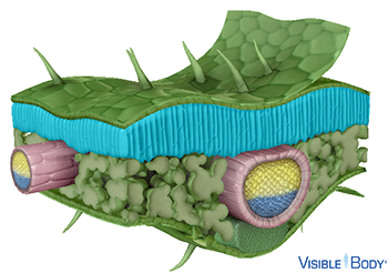
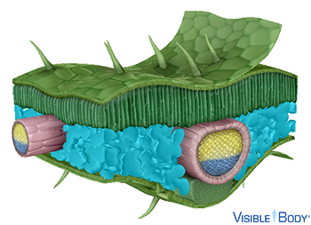
In vascular plants, the xylem and phloem form continuous tubes that carry water, nutrients, and other substances throughout the plant's roots, stem, and leaves. Negative pressure in the xylem moves water and dissolved minerals from the roots upward to the stem and leaves. The xylem contains several types of cells, including tracheids, vessel elements, parenchyma, and fibers. Tracheids and vessel elements are thick-walled cells that are dead at maturity, and they form side by side, connecting together to form tubes. See it in 3D!
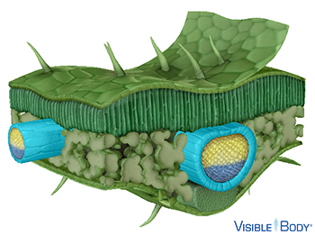
In vascular plants, the xylem and phloem form continuous tubes that carry water, nutrients, and other substances throughout the plant's roots, stem, and leaves. Positive hydrostatic pressure in the phloem moves dissolved sugars and organic compounds from the leaves downward to the stem and roots via a process called translocation. The phloem of monocots and dicots contains conducting cells (sieve elements) and companion cells. Conducting cells have thin walls, and they are alive in the mature plant, but they lack a nucleus and most other organelles. See it in 3D!
Dicot phloem also contains phloem parenchyma, fibers, and sclereids.
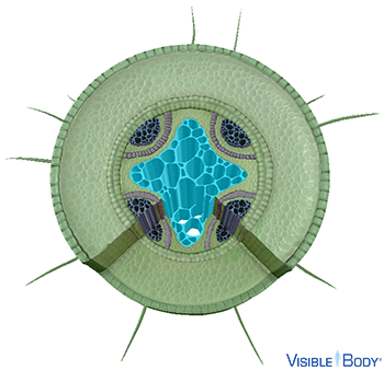
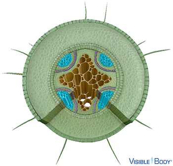
Dicot roots have a taproot structure, meaning they form a single thick root, with lateral branches, that grows deep into the soil. The ground tissue of dicot roots, primarily composed of parenchyma cells, surrounds the roots’ central vascular structures. See it in 3D!
The root's outer dermal tissue layer is the epidermis, a single layer of cells that protects the root and controls water and mineral absorption. See it in 3D!
Root hairs extend from the epidermis of monocot and dicot roots. Each root hair grows laterally as an extension of a trichoblast, a type of epidermal cell located in the root's maturation zone. Root hairs enhance the root's total surface area to maximize water and mineral absorption from the surrounding soil. See it in 3D!
Function: Their large surface area allows root hairs to efficiently absorb water via osmosis and minerals via diffusion. Their large central vacuoles allow them to collect water and minerals, which are passed into the root.
The cortex is a ground tissue region found in monocot and dicot roots, located between the outer epidermis and the inner vascular structures. It is primarily composed of parenchyma cells. See it in 3D!
Function: The primary functions of the cortex are diffusing water, nutrients, and other substances into the inner vascular structures and storing starch.
The endodermis is a single layer of cylindrical parenchyma cells that separates the root's cortex and stele. Endodermal cells have thick walls, and their radial and transverse walls contain Casparian strips, waxy bands that prevent water from passing between the cells. See it in 3D!
Function: The endodermis regulates the flow of water, ions, and hormones into and out of the vascular structures within the stele.
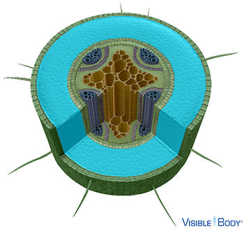
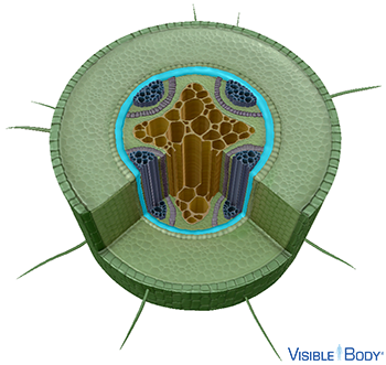
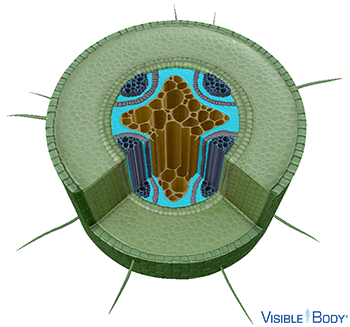
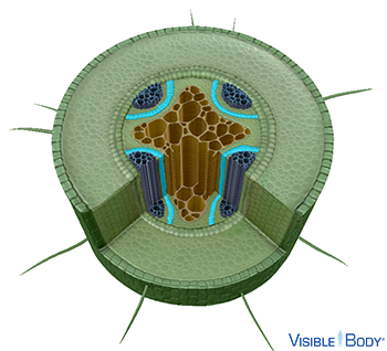
The stele (or vascular cylinder) is in the central region of the root, where the xylem and phloem develop. In dicot roots, the stele contains clusters of phloem arranged around the central xylem. Dicot steles contain an additional component, not present in monocot roots, called cambium. Dicot root steles do not contain pith. See it in 3D!
The pericycle is the outermost layer of the stele (or vascular cylinder). Although it is composed of nonvascular parenchyma or sclerenchyma cells, it is considered to be part of the vascular cylinder because it is produced by the procambium. See it in 3D!
Function: The pericycle supports the root, protects its vascular structures, stores nutrients, and facilitates root growth. Pericycle cells can divide and give rise to lateral roots in both monocots and dicots. In dicots, the pericycle also generates meristem cells that support secondary root growth and the cambium that produces xylem and phloem.
Parenchyma cells are the most abundant ground tissue cells, making up the majority of the cortex of dicot roots. Alive at maturity, they can divide to form new parenchyma cells. See it in 3D!
Function: They are large cells, with thin cellulose walls, that vary in shape depending on their function, which can include photosynthesis, respiration, gas exchange, and water and starch storage.
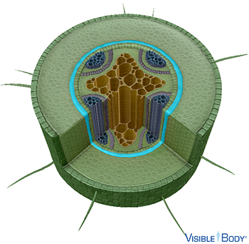
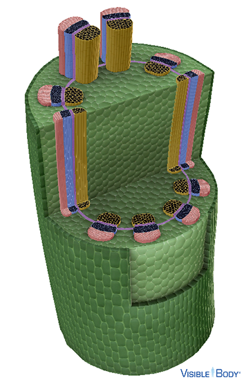
The steles of dicot roots contain a layer of meristem cells, called the cambium (or vascular cambium), located between the xylem and phloem. See it in 3D!
Function: These cells facilitate secondary growth, dividing to create new xylem and phloem cells, and thus widening the girth of the root.
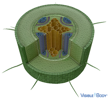
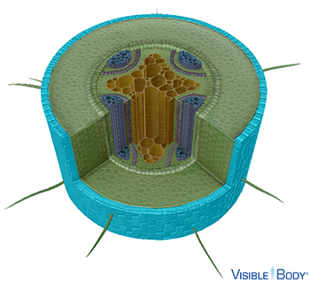
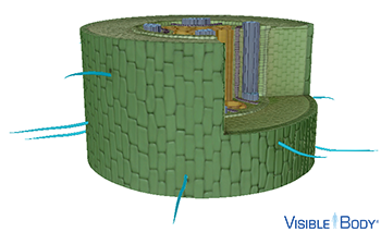
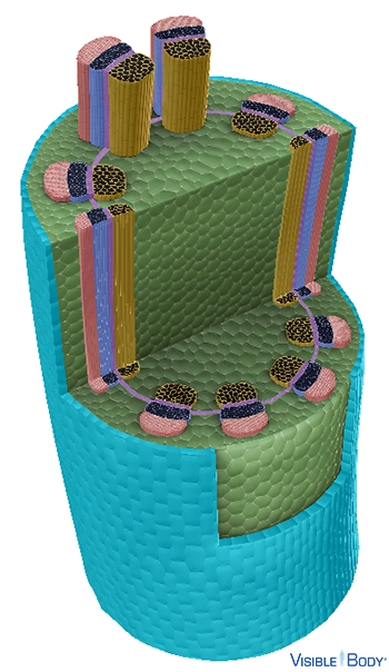
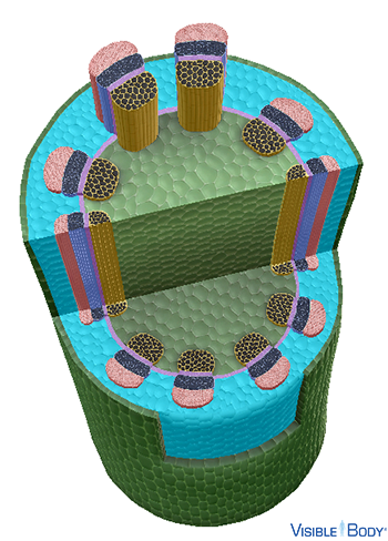
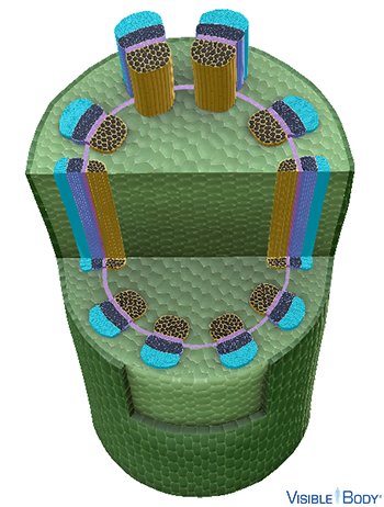
The majority of the dicot stem is composed of ground tissue, which primarily consists of parenchyma cells. Sclerenchyma and collenchyma cells are also found in regions that require extra strength. Dicot stems have a ring of vascular bundles, composed of xylem and phloem, that divide the ground tissue into the outer cortex and central pith. Dicot vascular bundles have an additional component, not present in monocot stems, called cambium. See it in 3D!
The stem's outer dermal tissue layer is the epidermis, a single layer of cells that prevents damage caused by sunlight, pathogens, and herbivores. In dicot stems, multicellular trichomes (hairs) extend from the epidermis. In woody dicot stems, the epidermis is replaced by periderm, composed of cork and other tissues. In some stems, the dermal cells secrete a waxy substance that forms a cuticle, a protective covering that helps the stem retain water. See it in 3D!
The hypodermis, composed of collenchyma cells in dicots, forms the stem's outer layer of ground tissue, located interior to the epidermis. See it in 3D!
Function: It provides structural support to the stem.
The cortex is a ground tissue region found in dicot stems, located between the outer epidermis and the inner vascular structures. It is primarily composed of parenchyma cells, and it may contain sclerenchyma and collenchyma cells in dicot stems. See it in 3D!
Function: The primary functions of the cortex are diffusing water, nutrients, and other substances into the inner vascular structures and storing starch. In the ground tissue, the cells are loosely arranged and there is space between them, which facilitates gas exchange between the stem and the surrounding air.
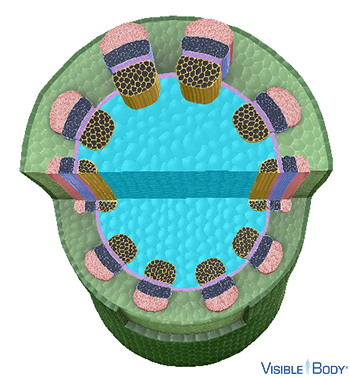
Located in the middle of dicot stems, the pith (or medulla) is composed of soft, spongy parenchyma cells with spaces between them. The pith is surrounded by a ring of vascular bundles, containing xylem and phloem. See it in 3D!
Function: Its primary function is storing water and nutrients and transporting them throughout the plant.
In dicot stems, sclerenchyma cells can be found in tissues where growth has stopped. There are two types of sclerenchyma cells, fibers and sclereids, which are dead at maturity and have thick, lignified cell walls. See it in 3D!
Function: Fibers are long, thin cells that provide strength to vascular bundles in stems, and sclereids are variably shaped cells that provide support for secondary phloem in dicots.
The vascular bundles of dicot stems contain a layer of meristem cells, called the cambium (or vascular cambium), located between the xylem and phloem. See it in 3D!
Function: These cells facilitate secondary growth, dividing to create new xylem and phloem cells, and thus widening the girth of the stem.
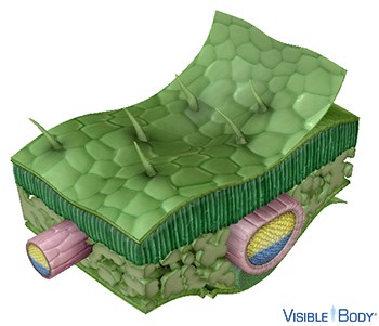
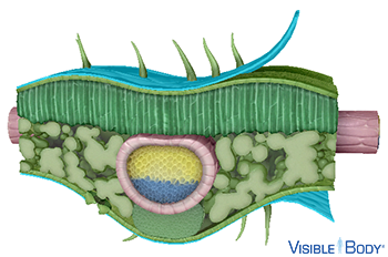
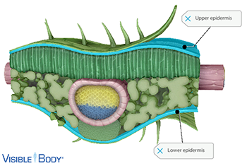
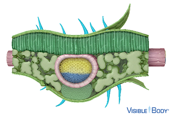
Dicot leaves have an upper and lower epidermis, and the lower epidermis contains several small pores, called stomata, which facilitate gas exchange and allow water vapor to exit the leaf. Having stomata only on its lower surface helps the dicot leaf conserve water while having most stomata open. The upper and lower epidermis are separated by ground tissue, called mesophyll, which contains parenchyma cells that carry out photosynthesis as well as sclerenchyma and collenchyma cells that provide structural support. In dicot leaves, the ground tissue is often arranged into two distinct types, the palisade and spongy mesophyll. The dicot leaf's vascular structures form net-like veins that are connected to the stem's vascular structures. See it in 3D!
The leaf's dermal cells produce and secrete a waxy substance, composed of lipid polymers, that forms a protective covering called the cuticle. See it in 3D!
Function: It acts as a barrier that prevents water loss via evaporation and protects the leaf from light damage and invading pathogens, such as viruses, bacteria, and fungi.
Leaves have two outer dermal tissue layers, the upper and lower epidermis, each composed of a single layer of cells that prevents damage caused by sunlight, pathogens, and herbivores. See it in 3D!
Most dicot leaves have trichomes, hairs that extend from the epidermal cells of the upper and lower epidermis. Trichomes can also be present on dicot stems, as well as monocot leaves and stems. Depending on the type of plant, trichomes can be unicellular or multicellular, branched or unbranched, and glandular or nonglandular. See it in 3D!
Function: They protect the plant from damage, which can be caused by herbivores, UV light, and extreme temperatures.
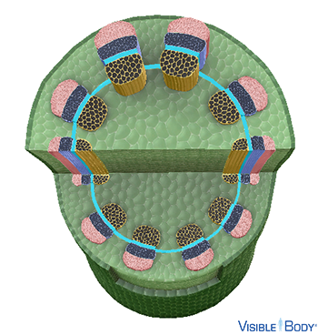
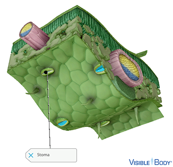
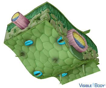
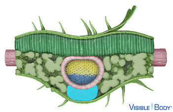
Stomata (singular: stoma) are small epidermal pores that allow gases and water vapor to move between the interior leaf structures and the surrounding air. In dicot leaves, the lower epidermis contains several stomata. Having stomata only on its lower surface helps the dicot leaf conserve water while having most stomata open. Note: Stomata are also present in photosynthetic stems. See it in 3D!
Each stoma is surrounded by two guard cells, specialized epidermal parenchyma cells that open and close the pore. See it in 3D!
Function: These cells regulate photosynthesis and respiration, facilitating gas exchange while open and preventing excess water loss while closed.
In dicot stems and leaves, collenchyma cells can be found in tissues where growth is occurring, such as near vascular cambium. Alive at maturity, these elongated cells have thick cellulose and pectin walls. See it in 3D!
Function: They provide flexible structural support.
In monocot and dicot leaves, sclerenchyma cells can be found in tissues where growth has stopped. There are two types of sclerenchyma cells, fibers and sclereids, which are dead at maturity and have thick, lignified cell walls. See it in 3D!
Unlike most monocot leaves, which have only one type of mesophyll, the ground tissue of dicot leaves is often arranged into two distinct types, the palisade and spongy mesophyll. In the palisade mesophyll, parenchyma cells are tightly packed, and their shape is usually polyhedral, elongated, or lobed. See it in 3D!
Function: This structure and the presence of chloroplasts facilitate photosynthesis.
In the spongy mesophyll, parenchyma cells are more loosely arranged, with spaces between them, and their shape tends to be spherical or stellate. See it in 3D!
Function: This structure allows the leaf to receive carbon dioxide from the air and to release oxygen and water vapor into the air.
A bundle sheath surrounds each vascular bundle, or vein, in monocot and dicot leaves. It consists of tightly arranged parenchyma cells in dicots. See it in 3D!
Function:These cells protect the vascular structures and play a role in photosynthesis.