
Whereas somatic cells in plants and animals divide via mitosis, sex cells (gametes), such as sperm and egg cells, form via a process called meiosis.
Germ cells in multicellular eukaryotic organisms divide via meiosis to form gametes. In humans, these germ cells are primary oocytes and primary spermatocytes.
What’s so special about gametes? Gametes aren’t exact copies of their parent cell like somatic cells. They are haploid cells, or cells with just one set of chromosomes, whereas somatic cells in most animals (including humans) are diploid, with two sets of chromosomes.
This is important because sexual reproduction requires fertilization, the union of cells from two different individuals, which forms a (diploid) zygote with one set of chromosomes from each parent. This means that for diploid organisms, gametes must contain only one copy of each chromosome.
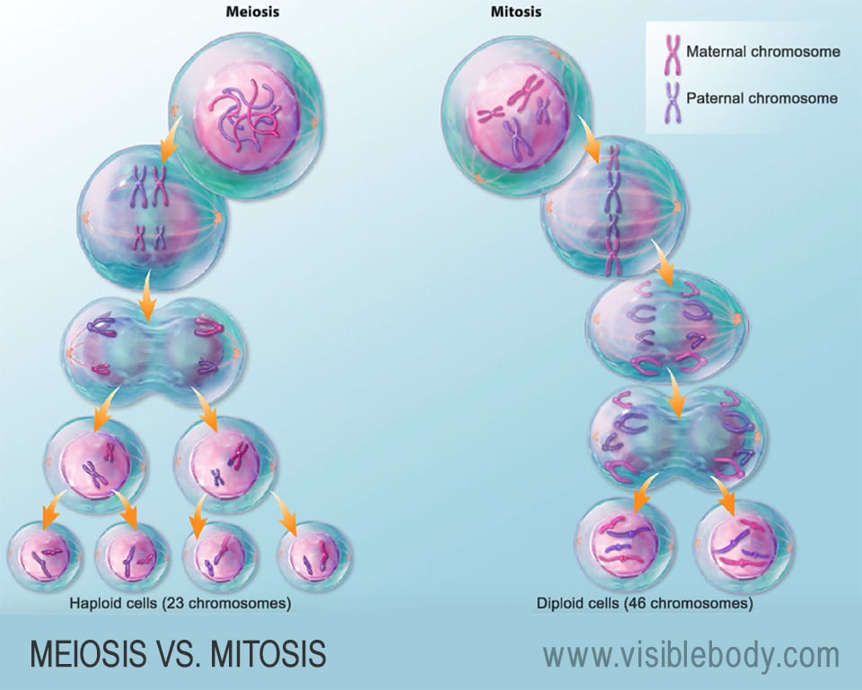
Let’s consider humans as an example. Our diploid somatic cells and germ cells have 46 chromosomes total. This means our haploid gametes — sperm and egg cells — each have only one set of 23 chromosomes. So a human zygote has one set of 23 chromosomes from each parent, for a total of 46. We can therefore say that our full set of 46 chromosomes consists of 23 pairs of homologous chromosomes — that is, chromosomes that are the same size and contain the same types of genes.
So how does cell division work when you start with a diploid cell and need to end with haploid cells? Meiosis!
Like somatic cells, germ cells spend most of their time in the G, or nonreplicative, phases of interphase of the cell cycle. During the S phase of interphase, their DNA replicates in preparation for meiosis.
Meiosis has two divisions — aptly named meiosis I and meiosis II — and each of these has five stages. You might recognize the names of these stages from studying mitosis: prophase, prometaphase, metaphase, anaphase, and telophase. However, pay close attention to the chromosomes during the phases of meiosis, and you’ll see just how different meiosis is from mitosis.
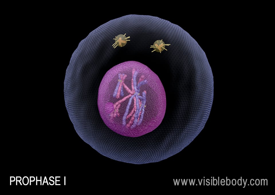
Meiosis begins with duplicated chromosomes, just like mitosis. Because they’re duplicated, each chromosome has two sister chromatids, joined by a centromere.
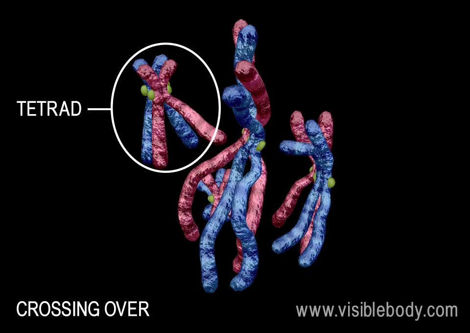
Remember the concept of homologous pairs we discussed earlier? They're going to be very important here. During prophase I, the duplicated chromatin condenses to form chromosomes, just like during mitosis. However, in meiosis (but not mitosis), these chromosomes organize into their homologous pairs, forming tetrads, so named because each chromosome has two sister chromatids, for a total of four chromatids in the tetrad.
Within each tetrad, non-sister homologous chromatids swap segments in a process called crossing over or recombination. This is where we start to see genetic variation. Chromatids where sections have been exchanged are referred to as recombinant chromosomes.
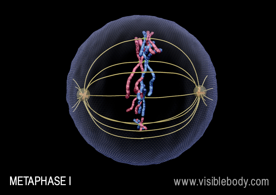
During prometaphase I, the nuclear envelope dissolves and spindle fibers attach to the centromeres and start to move the tetrads towards the center of the cell.
Metaphase I sees the tetrads line up at the center of the cell. The arrangement of homologous pairs during this stage is random — meaning that it’s random which duplicated chromosome from each homologous pair will go to which daughter cell. This is called independent assortment and is another source of genetic variation during meiosis. Independent assortment is also one of Mendel’s Laws of Inheritance: when alleles are sorted into gametes, genes don’t influence one another and every combination of alleles for every gene is equally likely to occur. Metaphase I shows how this law plays out biologically.
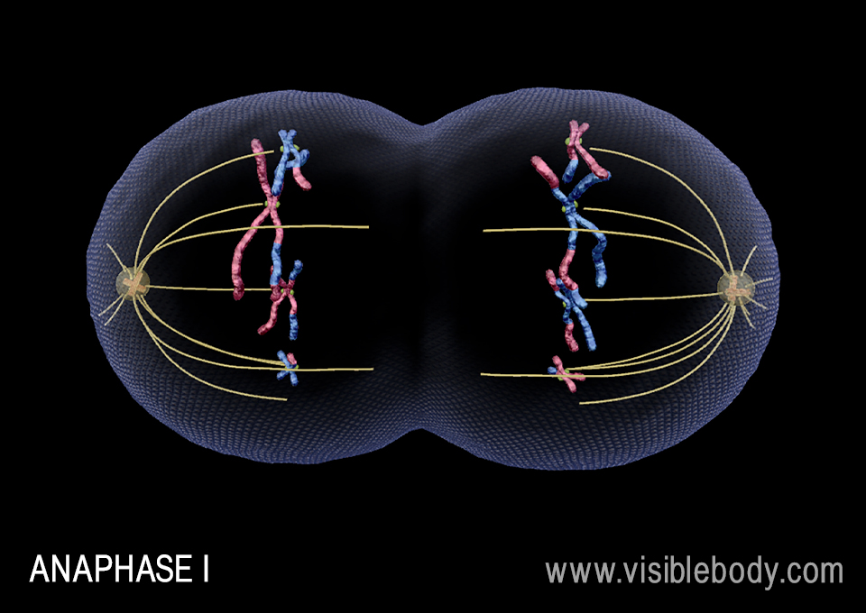
During anaphase I, the cell elongates as the spindle fibers pull the tetrads apart, bringing homologous chromosomes towards opposite ends of the cell. This part of the process is where we see Mendel’s Law of Segregation – which states that genes must segregate into gametes such that there is an equal chance of inheriting either version of the gene -- in action.
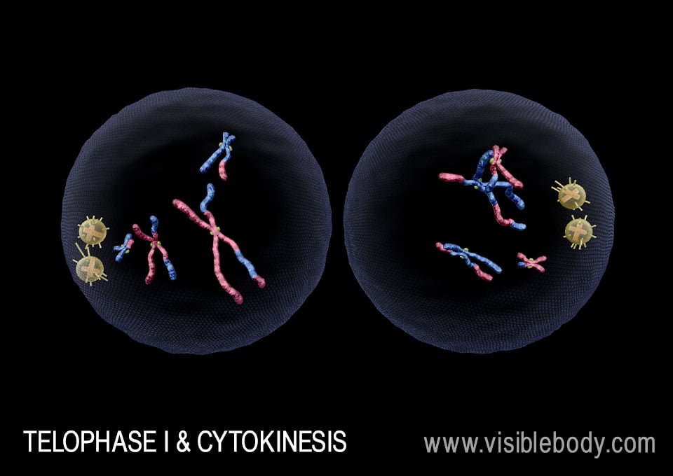
During telophase I, the chromosomes have reached opposite ends of the cell and the cleavage furrow forms. Then, cytokinesis splits the cell into two daughter cells. Note that each of these daughter cells is technically haploid — that is, it only has 23 chromosomes, even though they are duplicated chromosomes.
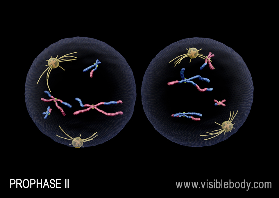
Next, each daughter cell from meiosis I undergoes division during meiosis II. Meiosis II looks a lot more like mitosis than meiosis I does. The sister chromatids in each cell will split to produce four genetically unique gametes. Some cells “wait” to complete meiosis II after meiosis I, spending time in a phase called interkinesis.
When it’s time for meiosis II, here’s how it goes. First, the spindle begins to form during prophase II. Then, during prometaphase II, spindle fibers attach to the chromosomes and start to move them towards the center of the cell.
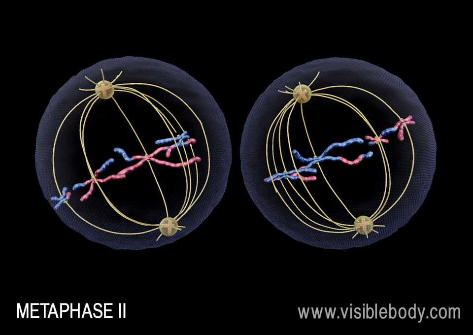
The spindle fibers line the chromosomes up at the center of the cell during metaphase II.
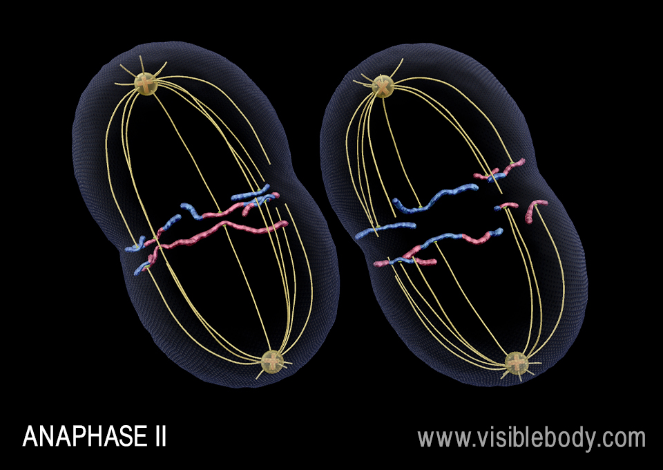
During anaphase II, the cell elongates and spindle fibers pull the sister chromatids apart, beginning to move them towards opposite ends of the cell.
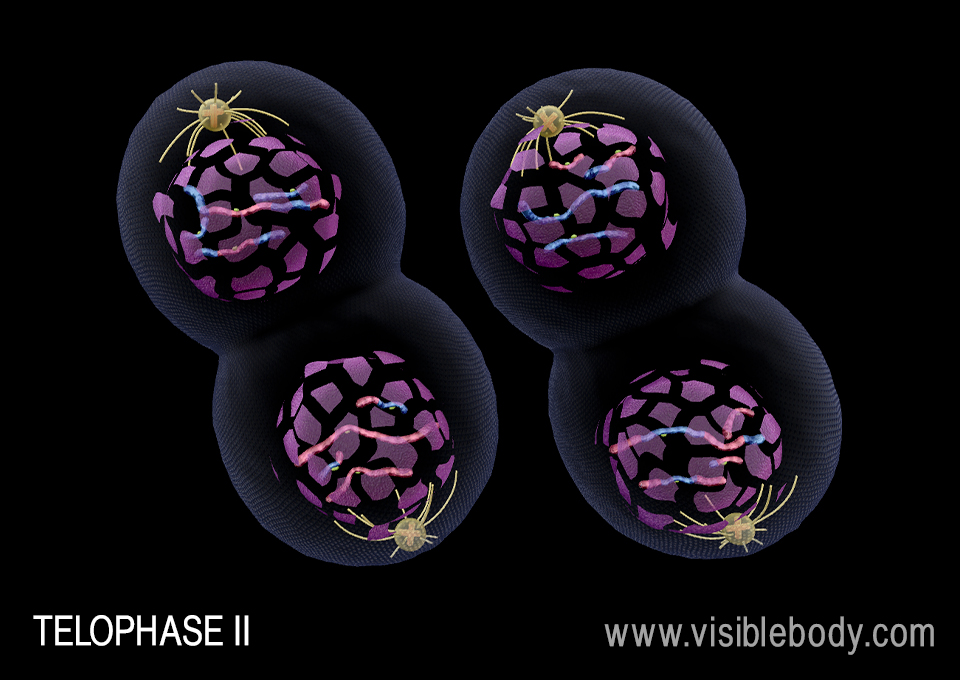
The cleavage furrow starts to develop in each cell during telophase II. In addition, a nuclear envelope forms around the chromosomes in each side of each cell, and a nucleolus forms in each new nucleus.
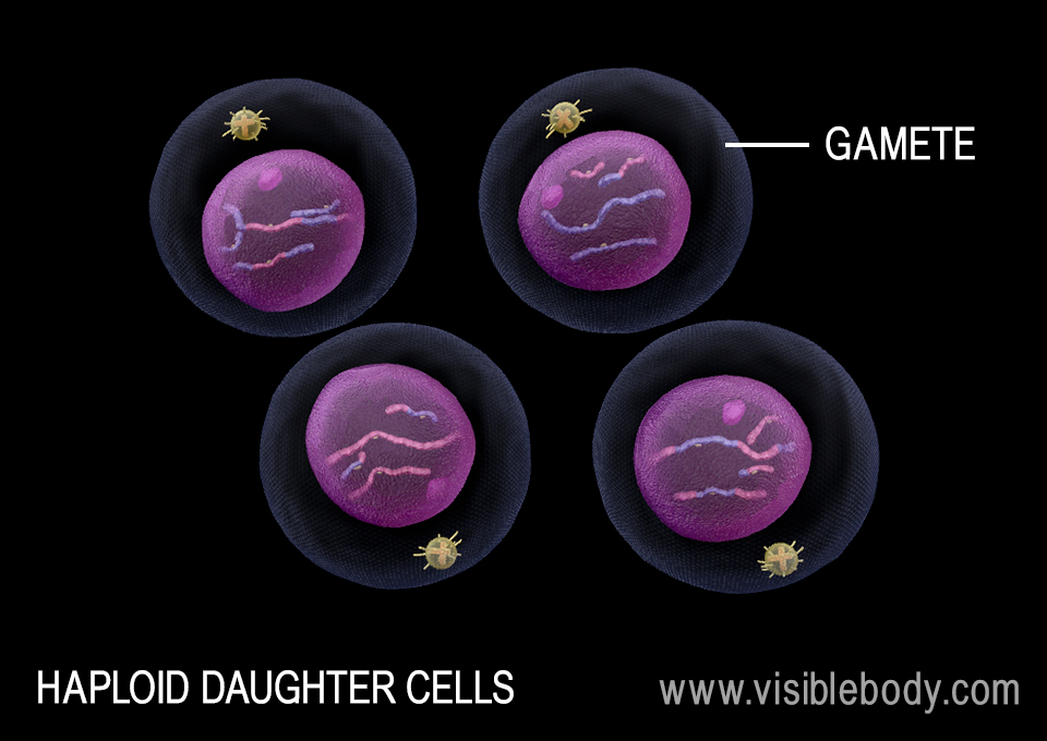
After cytokinesis, the result is four genetically unique daughter cells — gametes — each with only a single set of chromosomes. For humans, this means that gametes have 23 chromosomes.
Meiosis is a complex process! If you need a quick review, here’s a handy chart to summarize the steps of the cell cycle and meiosis:
| Interphase | G1 (growth, protein synthesis, organelle production) G0 (rest phase) S (DNA replication) G2 (protein synthesis, preparation for meiosis |
| Meiosis I | Prophase I (chromosomes condense, homologous pairs stay together and undergo crossing over) Prometaphase I (spindle fibers attach to centromeres, start to pull tetrads towards the center of the cell) Metaphase I (tetrads line up, independent assortment) Anaphase (homologous chromosomes separate) Telophase I (daughter cells begin to separate, followed by cytokinesis) |
| Meiosis II | Prophase II (spindle fibers form) Prometaphase II (spindle fibers start to pull chromosomes towards the center of the cell) Metaphase II (Spindle fibers line the chromosomes up at the center of the cell) Anaphase II (Spindle fibers pull the sister chromatids apart) Telophase II (A nuclear envelope and nucleolus forms for each new nucleus) |
| Cytokinesis | The products of meiosis II become four separate haploid gametes. |
