

Visible Biology’s Visual Guide to Pig Anatomy
In the biology classroom, students can learn a lot from examining pig anatomy. The pig’s major systems are similar to humans’, and students think critically as they compare and contrast these structures.
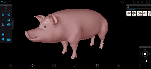
Launched in March 2023, Visible Biology’s interactive 3D pig model illustrates pig anatomy without lab prep or cleanup!
Visible Biology is available through Courseware, Visible Body’s LMS that can be used as standalone or integrated with Canvas or Blackboard. Through Courseware, instructors can use groundbreaking 3D models of biological concepts to create Flashcards, interactive Tours, dissection quizzes, and more!
Let’s take a look at the pig’s systems and structures.
Integumentary and muscle systems
A pig’s skin, aka integumentary system, is made up of two layers: the dermis and the epidermis. The dermis is on the interior while the epidermis is on the exterior. The dermis and epidermis contain nerve endings, hair follicles, and sweat glands.
Ever wonder why pigs love to roll in mud? Pigs’ sweat glands aren’t very good at keeping them cool, so mud helps pigs regulate their body temperature.
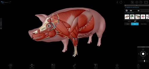
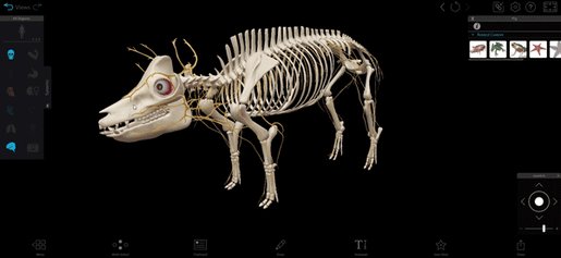
Pig model from Visible Biology.
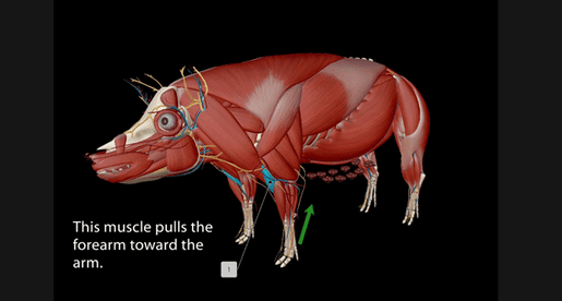
The pig’s head includes muscles like the masseter muscles (which move the jaw and help the pig chew) and the sternomastoid muscle (which helps the pig move its head). The brachiocephalic muscles stretch from the head to the front legs, helping the back muscles move the legs. Chest muscles like the pectoralis major and shoulder muscles like the deltoids also help move the front legs.
In the pig’s front legs, the biceps brachii and brachialis muscles pull the forearm toward the arm while the triceps brachii muscles extend the arm.
 Flashcards are a great study tool! You can make custom Flashcards with Visible Biology.
Let’s move on to the pig’s thorax and abdomen. The diaphragm is crucial for a pig’s breathing. When it contracts, air moves into the lungs, and when it relaxes, air moves out. The intercostal muscles also help the pig breathe by lifting and lowering the ribs; this is important because when the diaphragm contracts, the thoracic cavity expands. Take a deep breath—as you inhale, you can feel your own thoracic cavity expand thanks to your intercostal muscles.
Last but not least, let’s look at the muscles on the hind end of the pig. The gluteus minimus and gluteus maximus help the pig move its thigh: the gluteus minimus moves the thigh away from the body (abduction) and the gluteus maximus extends and externally rotates the thigh. The semitendinosus and semimembranosus muscles help move the lower leg and internally rotate the thigh. The biceps femoris and rectus femoris flex and extend the leg, respectively, and the gracilis muscles help move the legs towards the pig’s body.
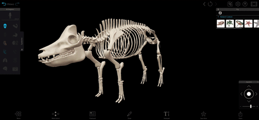
Skeletal system
The cranial bones in the skull protect the brain while the facial bones protect the respiratory and digestive organs housed in the skull. The mandible bone makes up the pig’s lower jaw and is the force behind chewing. Pigs also have a bone called the hyoid that supports the weight of the tongue.
The vertebrae protect the spinal cord and support the weight of the abdomen, and the caudal vertebrae at the end of the vertebral column form the pig’s tailbone. Exactly how many vertebrae does a pig have? That depends on the species.
Vertebrae and tail. GIF from Visible Biology.
The thoracic vertebrae articulate with the ribs. Pigs have 14 pairs of ribs, which protect the organs inside. The sternum protects the pig’s heart.
Let’s move to the forelimbs. The scapula makes up the shoulder girdle, and the elbow joint is formed by the humerus, radius, and ulna. The radius and ulna articulate with notches on the humerus.
Eight carpal bones make up the wrist. The metacarpals extend from the carpals to the phalanges, the bones in the digits. Each hoof has four digits, and each digit has three phalanges.
Next, let’s look at the hindlimbs. The pelvic girdle is made up of fused bones, and it articulates with the femur. The femur articulates with the tibia and fibula to form the knee joint, which is protected by the patella, aka the kneebone. The tibia articulates with the tarsals, aka the ankle bones. Just like with the front legs, the tarsals connect to the phalanges.
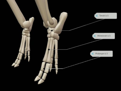
Hind leg ankle and foot anatomy with Visible Biology.
Nervous system
The central nervous system of the pig is made up of the brain and the spinal cord.
The brain is made of gray and white matter tissue and is covered by meninges. Meninges are layers of protective membranes.
GIF from Visible Biology.
Here are some of the structures in the pig’s brain:
- The cerebrum, which is responsible for voluntary muscle movements and the processing of sensory information, thoughts, and memory.
- The cerebellum, which is responsible for coordination and smooth movement.
- The brainstem (made up of the pons and medulla oblongata), which helps regulate involuntary functions. It connects to the spinal cord.
In addition to relaying messages to the brain, the spinal cord also processes reflexes.
The peripheral nervous system is a system of complex nerve networks that relay messages between the brain, spinal cord, and body. The pig’s 12 pairs of cranial nerves relay information between the brain and the sensory organs in the head and upper body, and around 30 pairs of spinal nerves branch out of the spinal cord to reach the rest of the pig’s body.
Circulatory system
The pig’s heart has four chambers:
- The left atrium receives oxygenated blood from the pulmonary veins and empties that blood into the left ventricle
- The right atrium receives deoxygenated blood from the anterior and posterior venae cavae. It empties into the right ventricle
- The left ventricle pumps blood into the body
- The right ventricle pumps deoxygenated blood to the lungs
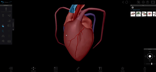
GIF from Visible Biology.
Oxygenated blood pumps out through the aorta, which branches off into arteries that supply the rest of the pig’s body with blood.
Deoxygenated blood moves from the heart to the lungs through the pulmonary artery and its branches. The pulmonary veins transport the newly oxygenated blood from the lungs to the heart.
Respiratory system
When a pig breathes, air enters the body through the nostrils and the mouth. It passes through into the pharynx and into the larynx, where the vocal cords are located. From the larynx, air passes through the trachea and into the lungs.
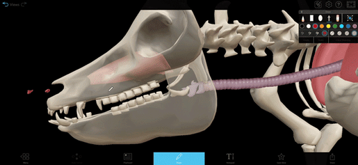
The Draw tool in Visible Biology.
The left lung has three lobes and the right lung has four. Airways called bronchi branch out from the trachea into the lungs, branching further into smaller airways called bronchioles. The bronchioles branch further into alveoli, where gas exchange occurs.
Digestive system
Digestion starts in the mouth, where the pig’s teeth grind down food and mix it with saliva, which contains digestive enzymes. The tongue helps the pig swallow and move food down the pharynx and into the esophagus.
From the esophagus, food enters the stomach. Glands in the stomach lining secrete juices that mix with food to break it down. Food travels through the pyloric sphincter into the small intestine, where it’s further digested and nutrients are absorbed. In the small intestine, more digestive juices and bile break the food down, and the remaining food passes into the large intestine, where it’s made into waste and travels through the rectum to the anus.
The liver, pancreas, and gallbladder are also part of the digestive system. In addition to filtering out toxins from the blood and making glycogen, the liver secretes bile into the small intestine; excess bile is stored in the gallbladder. The pancreas secretes digestive juices and produces glucagon and insulin, which regulate blood sugar levels.
.png?width=515&height=270&name=My%20project-1%20(16).png)
.jpg?width=515&height=270&name=My%20project%20(42).jpg)
Images from Visible Biology.
Excretory and reproductive systems
The excretory system is responsible for filtering blood and removing waste and excess water. The kidneys filter waste products like urea out of the blood; this process produces urine. Urine collects in the ducts and moves from the ureters to the bladder, where it’s stored before excretion through the urethra.
A female pig is called a sow and a male pig is called a boar—the Visible Biology model we’re showcasing is of a sow.
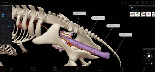
Image from Visible Biology.
Exterior female reproductive organs include the vulva (which contains the vaginal opening) and the urethral orifice (which is where waste is expelled). During fertilization, the cervix allows sperm to move from the vagina to the uterus. The ovaries produce hormones and cells that mature into eggs. During ovulation, some of the eggs are released into the oviducts, where they are fertilized. Then, the fertilized eggs move to the uterus.
A sow can give birth to up to twelve offspring at a time. The mammary glands and teats located on the sow’s abdomen produce milk for the piglets.
GIF from Visible Biology.
The boar’s gonads are the testes, located in the scrotum on the exterior of the body. The testes produce hormones and sperm cells. The testes connect to internal structures by the spermatic cord, which includes the vas deferens. Sperm moves through the two vas deferens, and the two vas deferens connect to form a single tube: the pelvic urethra. The pelvic urethra adds additional fluids to the sperm. The pelvic urethra continues into the penis, where it becomes the penile urethra, which transports both urine and semen.
Learn More
Check out the anatomy of the earthworm, sea star, and frog, Visible Biology’s other animal models:
- No Frog? No Prob! A 3D Visual Guide to Frog Anatomy
- Explore the Anatomy of Invertebrates with New Models!
Are you an instructor or administrator looking for resources for your course? Request an instructor code to try Visible Biology for yourself!
In addition to its 3D models, Visible Body supplies a rich library of free, premade teaching resources, and its attentive Customer Engagement team is available for one-on-one training sessions, so instructors who use Courseware are supported every step of the way. Visible Body offers premade Flashcard Decks, Tours, lab activities, and even premade courses that correlate to popular textbooks.
Read more blog posts for more teaching inspiration:
- Four Ways to Teach DNA Structure with Visible Biology
- How 3D Models Help Biology Students
- Free Lesson Plan: Diffusion and Osmosis with Visible Biology
- Reach More Students: Differentiated Instruction with Visible Body
Be sure to subscribe to the Visible Body Blog for more awesomeness!
Are you an instructor? We have award-winning 3D products and resources for your anatomy and physiology or biology course! Learn more here.
