No Frog? No Prob! A 3D Visual Guide to Frog Anatomy
There’s a reason frog dissection is the quintessential biology lab: examining frog anatomy teaches students about how organ systems function in complex organisms, drawing similarities between frog anatomy and their own.
If your classroom doesn’t have access to frog dissection specimens, Visible Body Suite's interactive 3D frog model can walk students through frog anatomy without prep or cleanup. It can also be a great way to prepare students for dissection, showing them structures they will see in 3D.
All of Visible Body's biology content is now exclusively available in VB Suite!
VB Suite is available through Courseware, Visible Body’s LMS that can be used as standalone or integrated with your LMS. Through Courseware, instructors can use groundbreaking 3D models of biological concepts to create Flashcards, interactive Tours, dissection quizzes, and more!
This ribbit-ing blog post will walk through the frog model in VB Suite and explore frog anatomy along the way. Let’s hop to it!
Integumentary System and Respiratory System
The frog’s skin (or integumentary system) performs protection and respiratory functions. The frog’s skin consists of two layers: the dermis (the deepest layer) and the epidermis (the outer layer). The skin is kept moist thanks to glands located throughout the dermis that secrete watery mucus. The frog’s skin is also semi-permeable, which means that it can absorb water and oxygen, and it can release water and carbon dioxide.
When the frog is in water, respiration takes place exclusively through the skin. On land, they take in air through the nostrils. Air then travels through the oral cavity to the glottis, a slit that opens into the larynx, where the air then moves into the lungs. The vocal cords are located inside the larynx. Frogs vocalize when air passes through the vocal cords. Ribbit ribbit.

GIF made using Tours in VB Suite.
Circulatory System
The frog’s heart is made of three chambers: the left atrium, the right atrium, and the ventricle. The skin and lungs provide oxygenated blood to the left atrium, and veins supply deoxygenated blood to the right atrium. The ventricle pumps blood through the aortas, where it’s then pushed through the body, and it pumps blood through the pulmonary vessels, where it moves to the lungs.
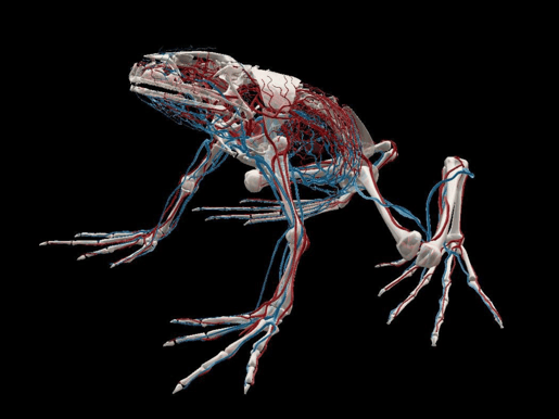 Image from VB Suite.
The system of veins and arteries throughout the frog’s body carries blood to and from the heart. The sinus venosus is attached to the right atrium and branches into the anterior vena cava and posterior vena cava which branch into other veins. The truncus arteriosus projects out of the ventricle and splits into three aortic arches which branch into other arteries.
The spleen removes old red blood cells and stores mature blood cells.
Skeletal System
Next, let’s move on to the skeletal system. The frog’s skull is broad and flat, encasing and protecting the brain, and it uses its mandible (lower jaw) and the maxillary teeth of its upper jaw to hold on to prey. The body and forelimbs are supported by a series of bones we might recognize from their human body equivalents: the suprascapula bones attach to the scapulae, which connect the clavicle bones to the sternum. The front limbs consist of a humerus and a radio-ulna, and the frog’s wrist bones, or carpals, articulate with the radio-ulna. Four metacarpals extend from the carpals and end with phalanges, aka toes.
 Frog bone Flashcard made with VB Suite.
The frog’s vertebral column protects the spinal cord and supports the head, and it consists of nine vertebrae. The last vertebra, the sacral vertebra, fuses with the pelvic girdle to form a structure called the urostyle. The pelvic bones articulate with the hind limbs, acting as a spring to help the frog jump. The frog’s back legs consist of a femur and a tibio-fibula bone. The frog’s ankles are made of two rows of tarsals
Five metatarsals extend out from the tarsals, and the phalanges extend from the metatarsals. The bones in a frog’s feet are elongated, giving the frog more surface area to push off from when it jumps.
Muscular System
A frog’s mouth and jaw are moved by the temporalis, submaxillary, masseter, and depressor mandibulae muscles, which work to elevate and lower the mandible and open and close its mouth.
The frog’s many forelimb and hindlimb muscles connect to the bones within the forelimbs and hindlimbs. The forelimb muscles extend and flex the bones in the forelimbs, and the hind limbs help the frog jump.
Between the forelimbs and the hindlimbs, the pectoralis, latissimus dorsi, and dorsalis scapulae help to move the forelimbs, the rectus abdominis flexes the vertebral column, and the longissimus dorsi extend the vertebral column and raise the frog’s head.
Digestive System
The frog’s retractable tongue catches prey and brings it into the oral cavity. The maxillary teeth and the vomerine teeth located on the roof of its mouth do not chew: a frog swallows its meal whole. Instead, the teeth hold prey still.
When the frog swallows, it passes food from the oral cavity and pharynx into the esophagus. The esophagus then passes the food into the stomach, where enzymes start to break down the food. Food then moves into the small intestine, where bile from the gallbladder and juices from the liver fully break down the food and nutrients are absorbed. Water is reabsorbed in the large intestine, and waste is excreted through the anus and into the cloaca. Waste exits the body through the cloaca, the cavity where products of the excretory and reproductive systems are released.
Fat bodies are the masses of fat that frogs contain in their body cavities. These stores of fat can be used when the frog has less food available, like during hibernation.
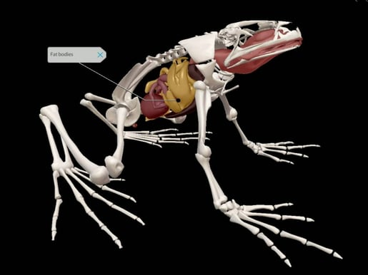 Image from VB Suite.
Excretory and Reproductive Systems
The adrenal glands are located ventral to the frog’s kidneys and secrete hormones that help with homeostasis. The kidneys filter waste products like urea out of the blood, producing urine. Urine flows through the ureters into the bladder, where it is stored before it’s excreted through the cloaca.
A male frog has testes that generate sperm cells and a female frog has ovaries that create eggs. Sperm and eggs are both released through the cloaca, and eggs are fertilized externally.
Nervous System
Let’s start with one of the frog’s most distinct features: those big ol’ eyes. Did you know that frogs have 360-degree vision? A frog’s eyes connect to the brain through optic nerves that stretch from the eye to the optic lobes in the forebrain. Transparent nictitating membranes act as third eyelids, protecting the eyes from debris and keeping them moist when the frog is out of water.

Image from VB Suite.
A frog’s ears consist of two main parts: the tympanic membrane and the inner ear. Tympanic membranes are located on either side of the head and transmit vibrations to the inner ear. Eustachian tubes connect the inner ear to the throat to equalize pressure in the inner ear when the frog is underwater.
The frog’s brain is divided into the forebrain, midbrain, and hindbrain.
The forebrain contains the…
- Olfactory lobe
- Cerebrum
- Diencephalon
The midbrain contains the…
- Optic lobes
- Nerve fibers connecting the diencephalon to the medulla oblongata
The hindbrain contains the…
- Medulla oblongata
- Cerebellum
The spinal cord extends from the medulla oblongata down the vertebral column. It processes the frog’s reflexes and conducts signals between the body and the brain.
The peripheral nervous system is typically made of 10 cranial nerves and 10 spinal nerves that carry sensory and motor signals between the brain and spinal cord.
Learn More
Are you green with envy after seeing that frog model? Are you an instructor or administrator looking for resources for your course? Request an instructor code to try Visible Body for yourself!
Visible Body supplies a rich library of free, premade teaching resources, and its attentive Customer Engagement team is available for one-on-one training sessions, so instructors who use Courseware are supported every step of the way. Visible Body offers premade Flashcard Decks, Tours, lab activities, and even premade courses that correlate to popular textbooks.
Read more and get some teaching inspiration on the blog!
- Four Ways to Teach DNA Structure with Visible Body Suite
- How 3D Models Help Biology Students
- Free Lesson Plan: Diffusion and Osmosis with Visible Body
Be sure to subscribe to the Visible Body Blog for more awesomeness!
Are you an instructor? We have award-winning 3D products and resources for your anatomy and physiology or biology course! Learn more here.

