How to Teach with Visible Body’s Dissectible Virtual Frog Model
With Visible Body Suite's 3D interactive frog model, students can learn about frog anatomy without any prep, cleanup, squeamishness, or clumsy scissor cuts.
Update: All of Visible Body's biology content is now exclusively available in VB Suite!
VB Suite is Visible Body’s visual guide to biological concepts and processes in addition to anatomy, physiology, and pathology. Its 3D model library covers prokaryotic and eukaryotic cells, monocot and dicot plant structure, energy, genetics, and blood cells—plus, students can explore vertebrate and invertebrate anatomy without extra equipment or cleanup. The app features four dissectible animal models: a frog, a pig, a sea star, and an earthworm.
In this blog post, you’ll find ideas for how to teach frog anatomy with VB Suite including how to run a virtual dissection lab.
Using this content
This blog post includes several features in Courseware, Visible Body's teaching and learning platform. All relevant content is already loaded into Courseware and ready to go!
If you don’t have access to Courseware yet, reach out to our team for a free instructor trial!
If you already have Courseware, follow these steps to add this folder to your course:
- Click on this link to add the lesson plan material to your Courseware account
- In this new course, click on the Bulk Editing tool, select the relevant folder(s) and click Copy. Choose the destination course and folder and copy.
- Navigate to your existing course, where you will find the content you copied. Use the bulk editing tool to edit release and due dates and publish. If you have Canvas deep integration set up, deploy to Canvas.
Once this lesson content is copied to your account, you can customize it to fit your class’s unique needs!
Lecture and review in 3D
With a library chock full of 3D models that illustrate biological concepts and processes, VB Suite is a game changer when it comes to lectures. The Tours feature makes for great lecture aids.

Tours are a series of Favorite views. GIF from VB Suite.
To create a Tour, start by positioning a model however you’d like—you can add structure tags, mark up the model with arrows and circles, and write text using the Notepad. Next, save that view to your Favorites. Next, save it to your Favorite views. Tours are collections of Favorite views—think about it like a presentation where you can reach in and move around the images on the slide to show more detail, answer students’ questions, and examine other structures before moving to the next slide.
You can make your own Tour, or you can use a premade one from our bank of free Tours!
When it’s time to review, try Flashcards. With a few easy clicks, you or your students can create custom Flashcards from the models. Making a Flashcard Deck is also a great assignment that challenges students to think critically about the takeaways from the day’s lesson.
Use structure tags or add your own prompts when creating Flashcards. GIF from VB Suite.
Just like with Tours, we also offer a free collection of Flashcard Decks, including one for frog functions. You can easily share Decks and Flashcards with your students—or vise versa—with a couple clicks.
The virtual dissection lab
Once students have a basic understanding of frog anatomy, it’s time to move on to a virtual frog dissection.

GIF from VB Suite.
Here are some tips and tricks to make the most out of the 3D frog model:
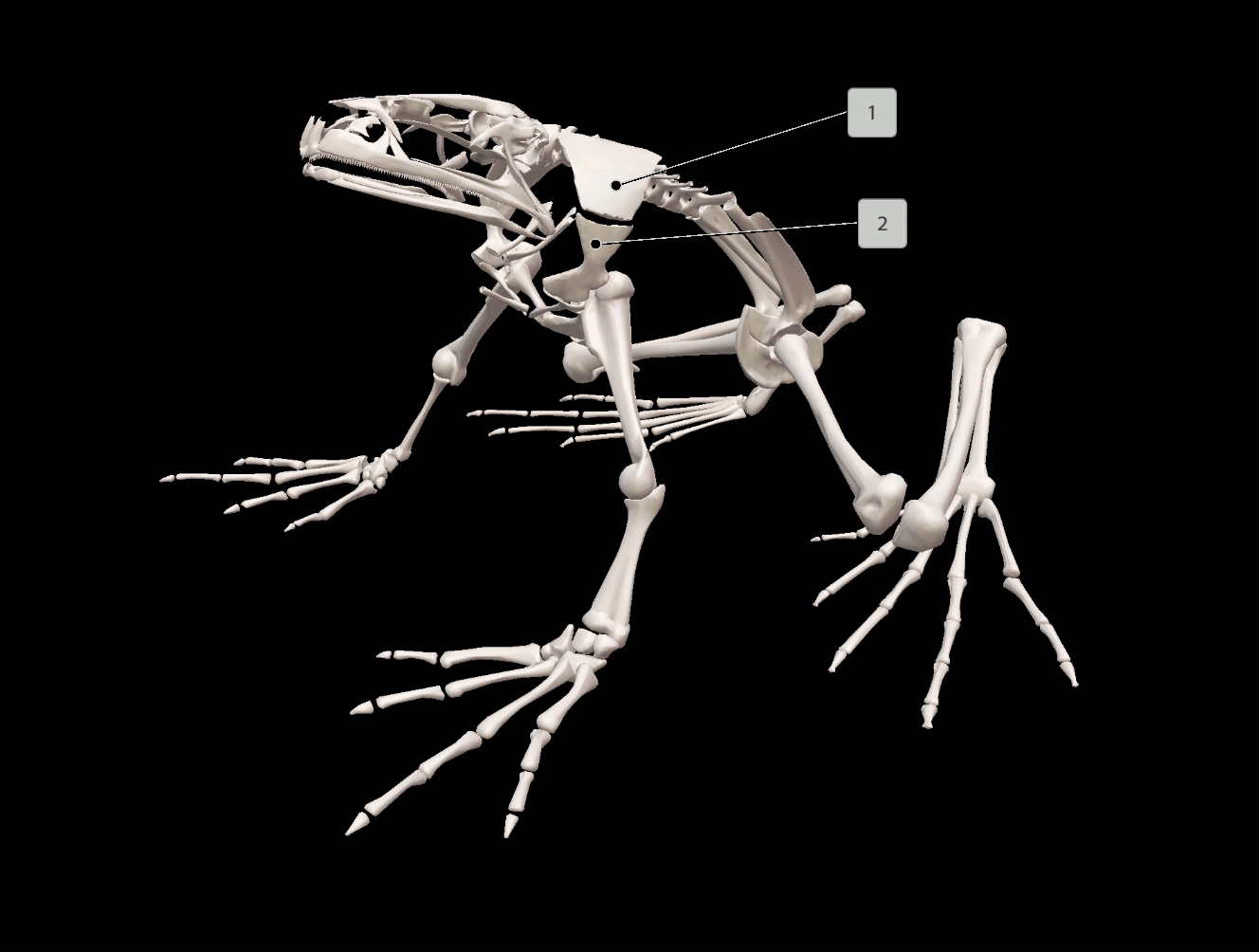
- The info box is full of great information that makes VB Suite just like a virtual textbook. When you select a structure, click on the definition icon to read about the structure and its context. Click on the pronunciation icon to hear how the name is pronounced—this is especially helpful for giving students the confidence to discuss anatomy. At any time, click on the information icon to read an overview of frogs.
- Keyboard shortcuts are your friend! VB Suite has many keyboard shortcuts to make manipulating models easier, and the list can be overwhelming at first. Before you work your way up to becoming a VB Suite super user, you can start small. There are two shortcuts that are particularly helpful for a virtual dissection. You can click on a structure and hit the H key to hide the structure and simulate a dissection, and you can click on a structure and hit the V key to fade the structure, making it appear transparent.
- The systems tray on the left side of the screen allows you to view or hide entire systems. This is helpful when you want to instantly hide the frog’s skin or if you want to look at a particular body system—for example, you could hide everything but the nervous system to see how the frog’s nerves all connect to the spinal cord.
With its free lab manuals, VB Suite has got your back when it comes to frog dissection labs and beyond. Designed alongside biology instructors, the free lab frog dissection manual was created for use with VB Suite’s frog model.
In the manual, your students will first find step-by-step instructions on accessing the model and answering the questions in the manual. The manual then leads students through background questions, activities on structure and function, and a step-by-step virtual dissection complete with follow up questions.
Assignment idea: Tour creation
In this assignment, students will create their own Tour, using VB Suite’s frog model to illustrate their answers. This assignment is designed to supplement the lab manual; once students have an understanding of frog anatomy, they will apply their knowledge. Through use of the Notepad and Draw tools, students will compose answers and provide visual aids to illustrate their points.
Students can complete this assignment individually or in groups, and this works for remote learning as well. It can easily be adapted for a homework assignment or a guided in-class exercise.
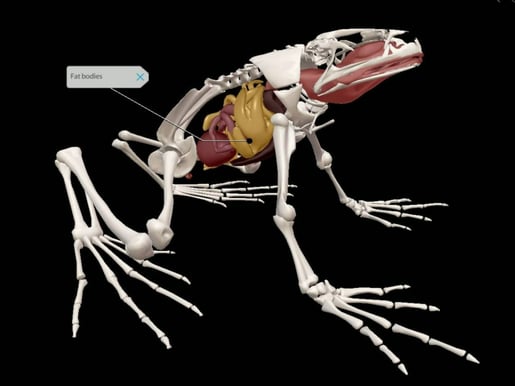
Image from VB Suite.
Assignment instructions
For this assignment, you will answer four prompts that ask you to apply what you have learned about frog anatomy. You will do this by creating a Tour in VB Suite and using the frog model to illustrate your answers.
How to create a Tour
A Tour is a collection of customized Favorite views—it’s what we used during the frog anatomy lecture.
Making Favorite views
Launch the VB Suite app and scroll down to the Animal Structure and Function category. Click on the frog model.
Using your mouse, move the model around. You can use the scroll wheel to zoom in and out. Just like the virtual dissection, you can remove structures by hiding or fading.
The Draw tool at the bottom allows you to draw freehand or add boxes, circles, and arrows to the model. The Notepad allows you to write your response to the prompt.
When you’re finished creating your view, click on Save View on the bottom toolbar and give your view a name, i.e., “Prompt 1.”
Making and sharing a Tour
To make a Tour, go back to the main menu. Click on the My Library tab at the top and then select Tours. Click Start to make a new Tour. Select your Favorite views in the order you want them to appear in your Tour, and then click Save.
To share your Tour, click on the three dots on your Tour’s thumbnail. Click Share Tour and copy the link it generates.
Turn in your assignment by pasting in the Tour link.
Assignment Prompts
You may need to make multiple Favorite views to answer each prompt.
Prompt #1: Think about where frogs live and what makes frogs suitable for their habitat. Pick three structures, and for each structure, explain in 3-5 sentences how it helps frogs live in their habitat.
Prompt #2: Next, consider how a frog interacts with its environment. In 2-4 sentences each, explain how the frog uses its senses of touch, taste, sight, smell, and hearing. Use the tag tool to tag the appropriate structures.
Prompt #3: Choose one body system (the systems tray on the left can help you view entire systems) and explain what it does. Using structure tags, point out three places where the system interacts with another body system. In 2-4 sentences each, name the other systems and explain how those structures interact.
Prompt #4: Find three frog anatomical structures that are analogous (functionally similar) to that of humans. Tag those structures and explain in 3-5 sentences each what makes these structures similar and different to those found in the human body.
Get VB Suite
You can request an instructor code to try Visible Body for yourself!
Visible Body supplies a rich library of free, premade teaching resources, and its attentive Customer Engagement team is available for one-on-one training sessions, so instructors who use Courseware are supported every step of the way. Visible Body offers premade Flashcard Decks, Tours, lab activities, and even premade courses that correlate to popular textbooks.
VB Suite is available through Courseware, Visible Body’s LMS that can be used as standalone or integrated with Canvas or Blackboard. Through Courseware, instructors can use groundbreaking 3D models of biological concepts to teach an interactive course.
Read more and get some teaching inspiration on the blog!
- How Can I Use Visible Body to Teach State Biology Standards?
- Four Ways to Teach DNA Structure with Visible Body Suite
- How 3D Models Help Biology Students
- Free Lesson Plan: Diffusion and Osmosis with Visible Body Suite
NGSS Standards
The above lesson ideas fit these Next Generation Science Standards (NGSS):
HS-LS1 From Molecules to Organisms: Structures and Processes. Students who demonstrate understanding can:
- Develop and use a model to illustrate the hierarchical organization of interacting systems that provide specific functions within multicellular organisms. (HS-LS1-2)
Did you know that the Visible Body Education Team has created a library of lesson plans and lab activities? Here's where you can find these resources, complete with NGSS standards.
Be sure to subscribe to the Visible Body Blog for more awesomeness!
Are you an instructor? We have award-winning 3D products and resources for your anatomy and physiology or biology course! Learn more here.

