5 Types of Medical Imaging
Here at Visible Body, it’s our goal to make the body, well, more visible! Our community has many medical and health sciences students, as well as folks who are just plain curious about the human body and how it works. Today on the Visible Body Blog, we’re going to talk about five imaging tests that health professionals use every day to gather information and pinpoint problems.
Let’s talk about x-rays, CT scans, MRIs, PET scans, and ultrasounds, how they work, and what they’re used for!
X-rays
X-rays are the most common diagnostic imaging technique, as well as the oldest.
An x-ray is a form of high-energy electromagnetic radiation that passes through most objects. During an x-ray test, x-rays travel through the body and are absorbed in different quantities by different organs. For example, the calcium content in bones means that they absorb x-rays better than other tissue, such as fat. An x-ray detector receives the x-rays after they pass through the body, generating an image that appears in shades of gray.
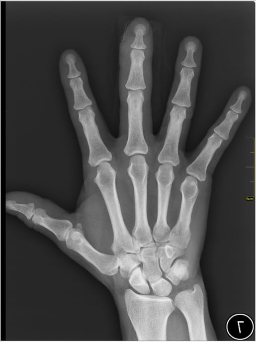
Hand x-ray image by Nevit Dilmen via Wikipedia Commons.
X-ray imaging is often the first imaging technique used when a patient has a problem, and they are used for a wide variety of purposes, ranging from looking for dental cavities to detecting cancer. X-rays are also used in other imaging techniques, like CT scans.
CT scan
CT stands for computerized tomography. You may have heard these scans called CAT scans, short for computerized axial tomography—both terms describe the same test, but CT scan is the more up-to-date term.
During a CT scan, the patient lies down on a table that slowly slides through the gantry, which is the circular part of the CT machine. Through the gantry, beams of x-rays are aimed at the body. The beams are rotated axially as the scan moves down the patient's body, and signals are sent to the machine’s computer. This generates cross-sectional images called tomographic images. These images can be put together to form a 3D image of the patient’s body, creating a more detailed view than normal x-rays.
X-rays are great at looking at bones and other dense structures, but x-ray images have a harder time picking up on soft tissues. That’s where contrast agents come in handy. Contrast agents are substances that are more visible on x-ray images—for example, iodinated contrast media (ICM), which are drugs that contain iodine and are delivered orally, intravenously, or rectally to make blood vessels and organs appear clearer on CT and x-ray scans.
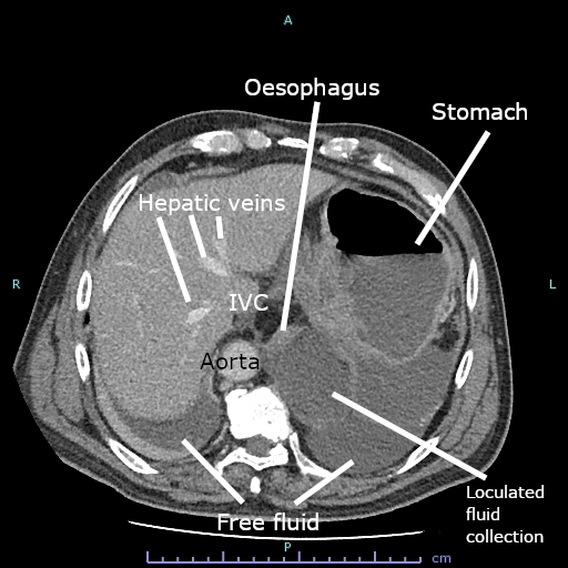
CT scan of fluid collection at the gastro-esophageal junction. Image by Cerevisae via Wikipedia Commons.
CT scans are used for a variety of purposes, such as:
- Diagnosing tumors
- Finding internal injuries such as internal bleeding
- Discovering heart disease
- Detecting complex bone fractures
MRI scan
MRI, short for magnetic resonance imaging, is a scan that uses both magnetic fields and radio waves.
The MRI scanner creates a strong magnetic field. Hydrogen ions in the human body spin around on their axes, and under the magnetic field, their axes all line up in the direction of the field. Most of the ions either face the feet or the head, effectively canceling each other out, but a few per million are not canceled out.
The machine then produces a radio frequency (RF) pulse that only affects hydrogen. When they are hit by the RF pulse, those leftover ions spin in a different direction. The RF pulse controls the frequency and direction of the hydrogen ions’ spinning.
Other magnets control exactly which part of the body is affected by the magnetic field—this can get as precise as “slices” a few millimeters in width.
When the RF pulse is turned off, the hydrogen ions release the energy they have absorbed from the pulses and return back to their alignment within the magnetic field. This produces radio waves that the MRI machine uses to generate images.
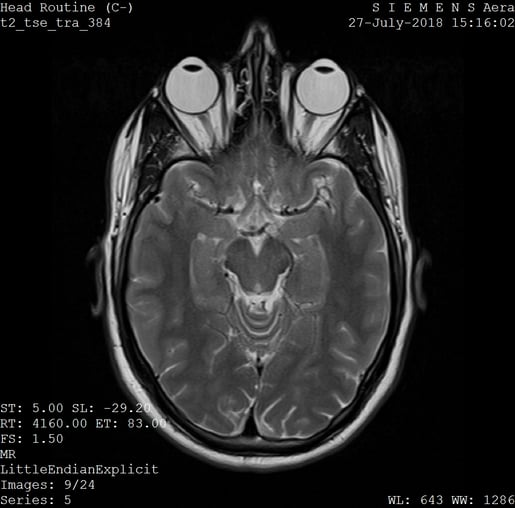
MRI scan of the head. Image by Ptrump16 via Wikipedia Commons.
MRI scans are often used to look at soft tissues and the nervous system. They can detect things like:
- Damage from a heart attack
- Brain injuries
- Cancer
- Multiple sclerosis
- Stroke
The strong magnetic fields of an MRI machine mean that metal objects cannot be brought into the room, and if a patient has metallic implants—for example, a pacemaker—this can complicate the scan process.
PET scan
PET is short for positron emission tomography. A PET scan measures cells’ metabolic activity, looking at biochemical changes to tissue and organs.
Before a PET scan, the patient must first take a radiopharmaceutical, or a drug containing a small amount of a radioactive substance. Radiopharmaceuticals can be ingested or administered intravenously.
One common radiopharmaceutical is fluorodeoxyglucose (FDG), which is similar in structure to glucose. FDG accumulates in malignant cells because they metabolize glucose very quickly. The PET scanner is able to find these malignant cells when it detects the gamma rays emitted by the radioactive substance.
Through analyzing the gamma rays emitted by the radiopharmaceutical, the PET scanner creates an image of the tissue or organ being scanned. The brightness of the image reflects how much radioactive material is present.
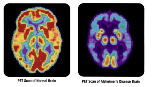 Images from the National Institutes of Health via Wikipedia Commons.
PET scans are mostly used to examine neurological and cardiovascular diseases as well as cancers. PET scans are able to detect most cancers before they can be seen on other tests, like CT and MRI.
Ultrasound
There are actually two categories of ultrasound: diagnostic and therapeutic. Therapeutic ultrasound uses sound waves to modify or destroy tissue, whereas diagnostic ultrasound uses sound waves to create images of the body. We’ll focus on diagnostic ultrasound for this blog post!
Transducers, or ultrasound probes, produce sound waves that cannot be heard by human ears. During an ultrasound test, gel is applied to the skin, and the ultrasound waves move from the transducer, through the gel, and into the body.
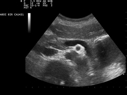
Ultrasound image of the pancreas. Image by Nevit Dilmen via Wikipedia Commons.
The sound waves hit an object and bounce back, creating an echo. The machine measures the echoes to determine the shape, size, and consistency of the object, generating a real-time image that appears on a screen.
Ultrasound is used for reasons like…
- Monitoring fetal growth and development
- Guiding a needle for a biopsy
- Examining the thyroid
- Diagnosing abdominal pain
Read more
Enjoyed this rundown on medical imaging? You may enjoy these other blog posts!
- Decoding the Heart: What Is an ECG?
- 4 Study Habits of Successful Med Students...Backed by Science!
- Can Augmented Reality Improve the Learning Experience of Future Healthcare Professionals? (2022)
- How Does Anesthesia Work?
Be sure to subscribe to the Visible Body Blog for more anatomy awesomeness!
Are you an instructor? We have award-winning 3D products and resources for your anatomy and physiology course! Learn more here.
Be sure to subscribe to the Visible Body Blog for more awesomeness!
Are you an instructor? We have award-winning 3D products and resources for your anatomy and physiology or biology course! Learn more here.

