Getting An Earful: The Anatomy Behind Hearing and Balance
Not only does the ear convert sound waves into nerve impulses, but it’s also responsible for our sense of equilibrium and movement. In fact, the ear is an organ so complex that it has more than one structure called a labyrinth!
The structures in the ear are divided into three categories: the outer ear, the middle ear, and the inner ear. Let’s go through them one by one and learn more about ear anatomy.
Outer ear
The outermost part of the ear is the auricle, also known as the external ear or the pinna. Made of cartilage covered in thin skin, the auricle amplifies sound and helps the brain determine the source of the sound.
The auricle has several parts—you might recognize some of the names from different types of ear piercings. Try using your own ear to follow along!
- The helix is the outer rim of the ear
- The antihelix is the prominence that runs parallel to the helix
- The scaphoid fossa is the depression at the top of the ear between the helix and antihelix, and the antihelical fold is the fold below the scaphoid fossa and between the helix and the antihelix
- The crus of the helix is the piece of cartilage above the ear canal, and the concha is the deep cavity between the crus of the helix and the antihelix
- The crura of antihelix are the next two pieces of cartilage that stick out above the crus of the helix, and the triangular fossa is the depression between the crura of antihelix
- The lobule (aka the earlobe) is the fleshy lowermost part of the ear and the only part of the outer auricle with no cartilage
- The antitragus is the ridge directly above the lobule, and the tragus is the pointy piece of cartilage that sticks out beside the concha and ear canal
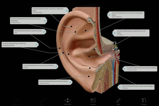
Image from Visible Body Suite.
The shape of the auricle is different for every person, and the brain knows how to locate sounds depending on how they bounce off the auricle’s unique structures. In fact, researchers have found that when you change someone’s auricle shape through silicone molds, they have a much harder time identifying where a sound is coming from—but after about a week, most of the participants adjusted to their artificial ear anatomy and were able to locate sound accurately again.
We’ve used the term ear canal, but let’s get a little more technical. The external acoustic meatus, also called the external auditory canal, is the skin-covered passageway that comes to an end at the tympanic membrane at the boundary of the outer and middle ear.
After bouncing around the auricle, sound is funneled through the external acoustic meatus and hits the tympanic membrane, also known as the eardrum. As you might have guessed, it’s drum-shaped! And just like a drum, sound causes the tympanic membrane to vibrate. Vibrations travel from the tympanic membrane to the middle ear.
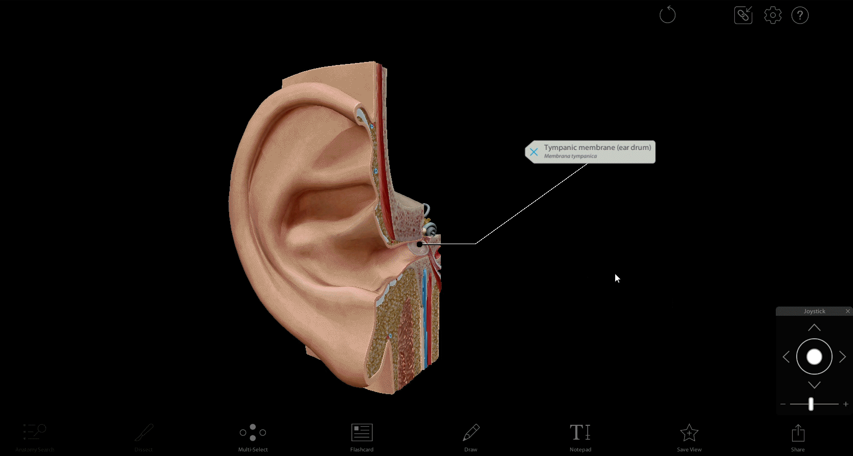
Sound travels through the external acoustic meatus and hits the tympanic membrane. GIF from Visible Body Suite.
The middle ear
The tympanic chamber of the middle ear is a hollow chamber filled with air. Attached to the tympanic chamber is the eustachian tube, the tube that connects the tympanic chamber to the nasopharynx of the upper respiratory system. When you swallow or yawn, the eustachian tube opens and equalizes the air pressure in the middle ear with the air pressure outside.
Ear pressure normalization is very important for scuba divers, because without it, they can experience barotrauma, which is an injury due to increased air or water pressure. When divers descend into the water, the change in depth can create a pressure difference between the chamber and the nasopharynx that yanks the tympanic membrane inward. This has the potential to cause the middle to fill with fluid as it tries to equalize the pressure. With severe barotrauma, the tympanic membrane can rupture, and a leak could even occur in the delicate structures of the inner ear. When diving, it’s important to be free of congestion so your eustachian tubes can function at their best and normalize the pressure in your ears.
In the tympanic chamber, there are three bones called the ossicles. The three ossicles are the malleus, incus, and stapes—also known as the hammer, anvil, and stirrup.
.jpg?width=2550&height=1700&name=My%20project%20(46).jpg)
The ossicles. Image from Visible Body Suite.
The malleus is attached to the tympanic membrane. The malleus receives vibrations from the tympanic membrane and transfers them to the incus, which transmits them to the stapes. Measuring at 0.1 inches, the stapes is the smallest bone in the human body!
From the stapes, vibrations move to the oval window, a membrane-covered opening between the stapes and the cochlea, located in the inner ear. The middle ear also contains the round window, another membrane-covered opening that allows the fluid in the cochlea to move with the vibrations transmitted through the ossicles.
The inner ear
The inner ear is responsible for a sense of balance in addition to hearing. The vestibulocochlear nerves are responsible for taking all of these signals to the brain. They have two branches, which we will talk about below.
The inner ear contains a system of tubes and sacs called the membranous labyrinth. This system is contained within a system of bony structures called the osseous labyrinth. The osseous labyrinth is divided into the vestibule, cochlea, and semicircular canals.
The vestibule is a chamber at the center of the osseous labyrinth. The semicircular canals are a series of three canals that connect to the vestibule in a total of five places. Each canal has an ampulla at each end, an expansion that contains fluid and hair cells. When the head rotates or moves, the fluid causes the hair cells to move. The vestibular nerves transmit impulses from the movement of the hair cells to the brain, giving the brain information about acceleration, deceleration, and equilibrium.
The cochlea is shaped like a spiral, and it connects to the vestibule, where it picks up on the vibrations sent through the oval window into the inner ear. When the oval window moves due to vibration, fluid pressure builds in the cochlea and activates the hair cells within. The cochlear nerves transmit the impulses to the brain.

The vestibule and cochlea. GIF from Visible Body Suite.
You may have heard of cochlear implants, which are electronic devices that can provide a sense of hearing to deaf or hard of hearing people. Where hearing aids amplify sounds, a cochlear implant picks up sound through a microphone, processes that sound, and then sends signals straight to the cochlear nerves, bypassing the other ear structures. People with cochlear implants who were not born deaf report that the sounds they experience through the implant are “mechanical” or “synthetic,” though most adjust to this over time.
Conclusion
The outer ear is used to funnel sound into the middle ear, where vibrations travel through the ossicles into the inner ear. In the inner ear, vibrations and shifts in acceleration, deceleration, and equilibrium are translated into nerve impulses through the movement of fluid and hair cells.
Thanks for hearing me out!
Read more on the Visible Body Blog:
Be sure to subscribe to the Visible Body Blog for more awesomeness!
Are you an instructor? We have award-winning 3D products and resources for your anatomy and physiology or biology course! Learn more here.

