At the Heart of Adaptation: A Comparative Anatomy Lesson Plan
Let’s get straight to the heart of the matter: here is a lesson plan based on the webinar below, delivered by the amazing Dr. Cindy Harley of Metropolitan State University.
By the end of this lesson, your students will understand the functions of the heart and why (some) animals have hearts. In addition to a hands-on activity that will compare the speed of circulation via diffusion versus a simulated heart, this lesson plan also contains links to premade labs that compare circulatory systems with the help of VB Suite's virtual, fully dissectible animal models: a sea star, an earthworm, a frog, and a pig.
Lecture: Three Basic Questions
First, let’s dive into three questions to cover in lecture.
What does a heart do?
A heart moves molecules around. It takes a long time to move through an animal's body just through diffusion, and animals have hearts so they can more efficiently get oxygen to their cells.
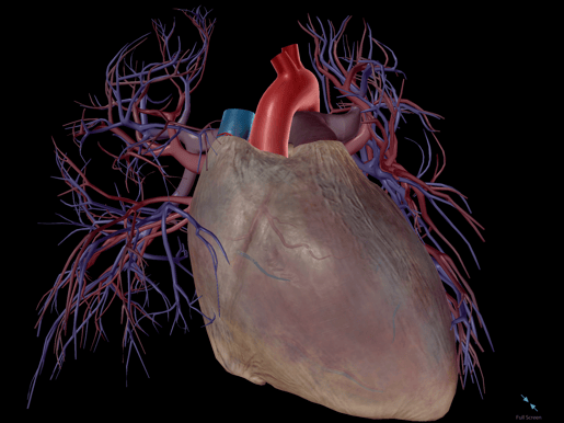
Beating human heart model in Visible Body Suite.
Why do some animals have hearts while others do not?
Let’s look at sponges as an illustrative example of why some animals don’t need hearts. Sponges do not have hearts—water flows through them easily, they don’t move and thus do not use up a lot of energy, and the body is so small that it's not a big deal to rely on diffusion.
We can also look at the sea star as an example. The sea star does not have a heart—in fact, sea stars don’t have blood at all. Instead, they have a water vascular system. Seawater enters the madreporite, and a system of canals keeps water flowing through their bodies.
Quickly remove body systems with the Systems Tray in VB Suite.
Do all animals’ hearts look the same?
Hearts don't have to be complicated! Some animals have hearts that are simpler than others. Simple hearts work for animals who don't need to move far and are low activity. These animals’ cells don't need as many nutrients. Animals who are more active need a more complex heart to get oxygen to their cells faster.
Take the fairy shrimp as an example. Although the fairy shrimp is small, it is so active that it can’t rely on diffusion; it needs a heart. The fairy shrimp has a simple heart that resembles a tube that runs down its back. When this tube squeezes, blood flows through the body.
Comparing Diffusion and the Heart
With two dishpans full of water, two pipettes, and some dye, you or your students can simulate the circulation of blood by the heart or through diffusion!
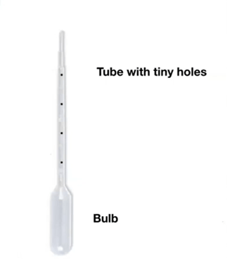
You will need to prepare two pipettes: one to illustrate diffusion and one to simulate a fairy shrimp heart.
For the diffusion pipette, simply fill it with dye.
For the fairy shrimp heart pipette, fill a pipette with dye and then poke holes along the side with a needle. Seal off the end of the pipette.
Screenshot from the Comparative Physiology of the Heart webinar.
Start by showing what the rate of diffusion might look like across an animal’s body. Place the pipette in one of the tubs of water and empty it in one squeeze. The dye will slowly diffuse into the water.
Next, simulate the fairy shrimp heart. Place the pipette in the water and squeeze it rhythmically to simulate a heartbeat. Soon, all the water will be colorfully dyed!
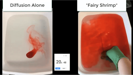
Screenshot from the Comparative Physiology of the Heart webinar.
Ten to twenty minutes later, the dye in the diffusion pan will not have disseminated yet, but the dye in the fairy shrimp heart pan should disseminate completely in under 30 seconds.
Lecture: Functions of the Heart
Humans’ hearts have two jobs: the first is to move deoxygenated blood to the lungs to pick up oxygen and the second is to move oxygenated blood to the tissues.
To explore heart functions, walk your students through the following examples.
Squids have three hearts. Two hearts send blood over the gills to become oxygenated and the other one (the systemic heart) moves the blood into the tissues.
Fish hearts work differently. A tube moves over the gill, oxygenating the blood. The oxygenated blood then moves to the tail. As the fish swims, the muscles in the tail apply pressure to the blood vessels, and blood is pumped back to the heart.
Fish have to keep moving to stay alive—as Dory says in Finding Nemo, "Just keep swimming."
Humans, on the other hand, require more complex hearts, containing a system of chambers and valves that keep blood flowing. Our hearts may not require physical activity to circulate blood, but it doesn’t mean they’re perfect for every situation.
Heart model in Visible Body Suite.
Crocodiles can stay underwater (not breathing) for long periods of time because of how their hearts are structured. Deoxygenated blood still has some oxygen in it, so when a crocodile is underwater, a structure in its heart will keep recirculating deoxygenated blood, bypassing the lungs. The body uses the remaining oxygen to function.
Human infants have a similar structure in their hearts! The lungs did not function while in utero, so the heart didn’t pump blood to the lungs for oxygenation.
Virtual Lab Activities: Comparing Hearts
VB Suite contains a visual guide to biological concepts and processes! The app features four animal models—a frog, a pig, a sea star, and an earthworm—that allow students to explore animal anatomy without any extra equipment or cleanup. In addition to vertebrate and invertebrate anatomy, its 3D model library covers prokaryotic and eukaryotic cells, monocot and dicot plant structure, energy, genetics, and blood cells.
Our education team has put together a free digital library of lab activities and lesson plans, including an animal circulatory comparison lab and several labs on human circulatory anatomy and function!
 Frog model in VB Suite.
You can also browse a bank of premade Flashcard Decks that include Decks on animal anatomy as well as human heart anatomy and circulation.
Looking for more free teaching resources? Check out these blog posts for some more lesson plan inspiration!
- A Heart Rate Lesson Plan with Visible Body Suite
- 4 Methods to Boost Student Engagement
- Comparing Animal Anatomy: A Visible Body Lesson Plan
- 4 Ideas for Teaching Earthworm Anatomy with Visible Body
- How to Teach with Visible Body's Dissectible Virtual Frog Model
- 5 Ways to Gamify Your A&P Classroom with Visible Body
To close things out, we'd like to thank Dr. Harley once again for delivering this fantastic webinar!
NGSS Standards
The above lesson plan fits these Next Generation Science Standards (NGSS):
HS-LS1 From Molecules to Organisms: Structures and Processes. Students who demonstrate understanding can:
- Develop and use a model to illustrate the hierarchical organization of interacting systems that provide specific functions within multicellular organisms. (HS-LS1-2)
Did you know that the Visible Body Education Team has created a library of lesson plans and lab activities? Here's where you can find these resources, complete with NGSS standards.
Be sure to subscribe to the Visible Body Blog for more anatomy awesomeness!
Are you an instructor? We have award-winning 3D products and resources for your anatomy and physiology course! Learn more here.

