The Benefits of Visible Learning: A Q&A with Bert Oppenheim
The Visible Body Blog was excited to sit down with Bert Oppenheim, Vice President of Creative Services here at Visible Body, to chat about medical accuracy, how learning in 3D can help students, and about Visible Biology, the company’s new biology product released in January 2022.
Check out what Bert had to say!
How does the Visible Body team ensure medical accuracy in their products?
All of our medical artists are academically trained, which means that each of our talented content creators have gone through rigorous schooling that combines art disciplines and biomedical science courses. This includes human anatomy courses with dissection along with other courses such as embryology, histology, and neuroanatomy. Not only do we have very qualified in-house artists checking accuracy, we also have many external reviewers including anatomists and professors/instructors who provide expert review and feedback for us.
What are gross anatomy and microanatomy views, and how do they help students gain a better understanding of the body?
Gross anatomy views are those that are large enough to see with our eyes. Much of what we have in our Human Anatomy Atlas are gross anatomy views. On the other hand, microanatomy deals more with the smaller structures and fine details—for example, the inner structures of the eye or the multiple different cellular layers of blood vessels. Many of these very small structures cannot be seen with our eyes but are rather identified with a microscope, so having these interactive 3D microanatomy models in our app helps students understand their connection to the larger gross anatomy views.
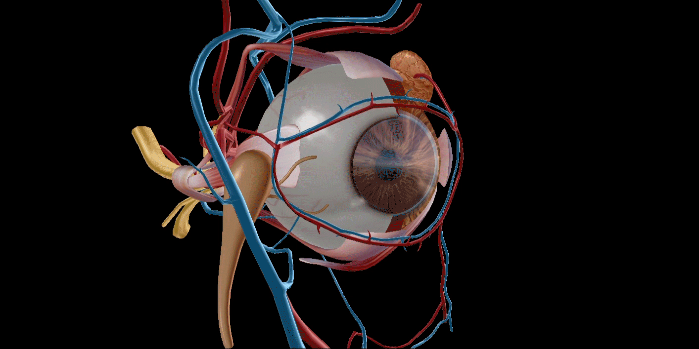
GIF from Human Anatomy Atlas 2022 +.
How does Visible Body help students visualize spatial and functional relationships?
This is one of my favorite topics to discuss. I think of the human body as a gigantic puzzle with thousands of individual pieces that can only fit together one way. A few examples include the differences between our lungs; our left lung is divided into 2 lobes while our right has 3 lobes. The left is slightly smaller because it allows room for our heart on that side of the body. When we think of our kidneys, the left kidney sits higher than the right kidney, the reason being that the right kidney has to accommodate the liver which takes up a lot of the space on that right side of the abdomen. If you looked at the position of the kidneys from the front, you wouldn’t imagine they sit so close to your spine unless of course you rotate our 3D anatomical model to the side. The ability to interact with our models provides that understanding and visualization for students learning anatomy.
In this short webinar, Bert looks at the differences between 2D and 3D learning.
How is pathology incorporated into Visible Body’s content?
In order to have a full understanding of the human body, you need to visualize what happens to our anatomy when things go wrong. Generally, we all begin learning about what happens in a healthy body, but when someone becomes sick and their organs become damaged, it’s important to have that comparison to understand how tissues and structures change from healthy to diseased. With our pathology models and animations, students can study 3D models of common diseases and conditions. A good example for teaching is comparing our normal microanatomy models of blood vessels with our 3D model of progression of atherosclerosis, plaque that builds up within the walls of our blood vessels.
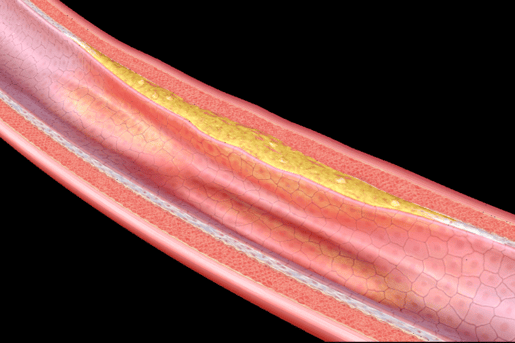
Early-stage atherosclerosis image from Physiology & Pathology.
Can 3D models help students build clinical knowledge?
Our 3D models can help build clinical knowledge for medical or nursing students who spend a lot of time with patients discussing their health. Clinical knowledge usually comes from learning how to diagnose and treat different problems, and our normal and pathology content helps to do that.
One example is the detailed model of the peritoneum. Medical students are required to learn the FAST (Focused Assessment with Sonography in Trauma) exam, which includes identifying the presence of fluid within the abdominal cavity, and our detailed model of the peritoneum is broken down into many structures and spaces where fluid generally shows up on a FAST exam.
What was it like to expand Visible Body’s content to include biology?
By definition, biology is the science of all living organisms, so it was an easy transition for us since for so many years we’ve been creating human anatomy 3D models and animations. With Visible Biology, we’re building models at the cellular level and developing animations of processes like photosynthesis or DNA replication.
I think all of us were ready to expand into biology. We heard from not only the market but many of our existing educational partners and professors that there is a strong desire and need to teach and offer biology content to their students. Visualizing interactive 3D biology models opens up a new way of visualizing the content compared to the static traditional pictures most have seen in the past through textbooks. It is a great feeling to see the development of our Visible Biology app come to the market after all the hard work from many different teams within Visible Body.
Last but not least, what’s your favorite view or animation?
Any of our microanatomy models, because most of that content is so very difficult to visualize on such a small scale. I think the level of detail that is captured within each model makes them special for me.
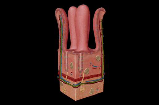
Image of intestinal villi from Human Anatomy Atlas 2022 +.
Wrap-up
A huge thanks to Bert Oppenheim for talking with us about learning in 3D!
To see our amazing 3D visuals for yourself, check out Human Anatomy Atlas 2022 +, where you can find full male and female body models, study tools, and more.
Be sure to subscribe to the Visible Body Blog for more anatomy awesomeness!
Are you an instructor? We have award-winning 3D products and resources for your anatomy and physiology course! Learn more here.

