Open Wide: A Guide to Dental Anatomy
The typical mouth has 32 teeth, and not only do they help us bite off and grind down food, but they even help us speak!
Today on the Visible Body Blog, we’re taking a closer look at teeth. We’ll look at dental vocabulary, the anatomy of teeth and gums, and the different categories of teeth.
Basic vocabulary
First, let’s go over some vocabulary that will help us talk about teeth.
Mandibular refers to the teeth on the lower jaw, also known as the mandible. Maxillary refers to the teeth on the upper jaw. The maxillae are the two facial bones that form the upper jaw.
.jpg?width=515&height=386&name=screenshot%20-%202023-10-02T092603.705%20(1).jpg)
Image from Visible Body Suite.
The anterior teeth are the twelve teeth in the front of the mouth, while the posterior teeth are the teeth in the back of the mouth. The anterior teeth consist of the incisors and canine teeth, and the posterior teeth consist of the premolars and molars. Loosely speaking, the anterior teeth are responsible for biting and the posterior teeth are responsible for grinding up food.
There are also several terms referring to the surfaces of the teeth:
- The lingual surface is that which is closest to the tongue.
- The buccal surface is the surface closest to the cheek.
- The distal surface is the surface away from the median line of the face.
- The mesial surface is the surface near the median line of the face.
- The incisal surfaces are the cutting edges of the anterior teeth.
- The occlusal surfaces are the chewing and tearing surfaces of the posterior teeth.
Anatomy of a tooth
Let’s start with an exterior look at tooth anatomy, starting with the root, the neck, and the crown!
The root of a tooth is composed mostly of dentin, which is covered in cementum. Cementum is a calcified substance that connects to the periodontal ligament (PDL), which is made of connective tissue that attaches the teeth’s roots to the bone. The fibers of the PDL also protect teeth from becoming too compressed during chewing. Different types of teeth have different numbers of roots—more on that later.
Between the root and the crown lies the neck of the tooth. This line between the root and crown is known as the cervical line or cementoenamel junction. The gingiva (aka the gums—we’ll talk more about the gingiva later) connects to the neck through gingival fibers.
Like the root, the crown is mostly made of dentin, but unlike the root, it’s covered in enamel instead of cementum.
In the image below, you can see the crown in blue, the neck in orange, and the root in light pink.
.jpg?width=515&height=386&name=screenshot%20-%202023-10-02T092922.956%20(1).jpg)
Image from Visible Body Suite.
Cusps are raised points of enamel. The number of cusps depends on the type of tooth. There are two main types of cusp: lingual cusps (located by the tongue) and buccal cusps (located near the cheek). Some teeth have cingula, which are ridges near the base of the crown.
Fossae are depressions, typically on the occlusal surface (the surface for chewing) of a tooth. Incisor and canine teeth have lingual fossae—their fossae are on the tongue-facing side of the tooth.
Toggling between cusps and fossae in Visible Body Suite.
Here’s a pro tip for Visible Body Suite users who want to get the most out of the app. To learn more about dental anatomy, click on any tooth and then go to the info box. Click on the colorful tooth icon, and you can access visualizations and information about cusps, fossae, and surfaces of the teeth!
Tooth anatomy: an inside look
Now let’s take a look inside the tooth!
We’ve already talked about how most of the tooth is composed of dentin, and the dentin is covered in enamel in the crown and cementum at the root. Dentin is what surrounds the pulp cavity.
The pulp cavity is located in the center of the crown and extends into the roots of the tooth as pulp canals. Pulp’s main function is creating more dentin—when a tooth decays, the pulp works to replace the lost dentin. Pulp is made of connective tissue, and the blood vessels, nerves, and lymphatic vessels in that tissue give the teeth nutrients, sensation, and protection.
.jpg?width=515&height=386&name=screenshot%20-%202023-10-02T094451.133%20(1).jpg)
Molar crossection in Visible Body Suite.
At the base of the roots are the apical foramina, which are openings 0.3 to 0.4 millimeters in diameter. These openings allow nerves and blood vessels to enter the teeth and supply the pulp.
The gingiva
When you talk about teeth, you gotta talk about gums, too. The gingiva, also known as the gums, is a mucosal tissue that surrounds the alveolar bone and root(s) of the tooth. There are actually three types of gingivae:
- The marginal gingiva surrounds the tooth
- The attached gingiva connects to the periosteum (connective tissue) that surrounds the maxillae and mandibles
- The interdental gingiva provides a seal between the teeth
Types of teeth
Last but not least, let’s go over the types of teeth: the incisors, canines, premolars, and molars.
Incisors
Incisors have an incisal edge, which is a sharp edge that’s used for cutting food.
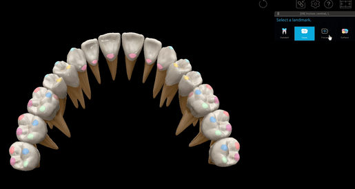
There are eight incisors in total: two maxillary central incisors, two maxillary lateral incisors, two mandibular central incisors, and two mandibular lateral incisors. The incisors are anterior teeth.
.jpg?width=515&height=386&name=screenshot%20-%202023-10-02T095058.510%20(1).jpg)
Image from Visible Body Suite.
The maxillary central incisors are your two front teeth! In addition to cutting food, they’re also used for speech. .
Some people are born without one or both maxillary lateral incisors—this is a condition called maxillary lateral incisor agenesis (MLIA). MLIA is the most common condition where permanent teeth are congenitally missing, accounting for 20% of those cases.
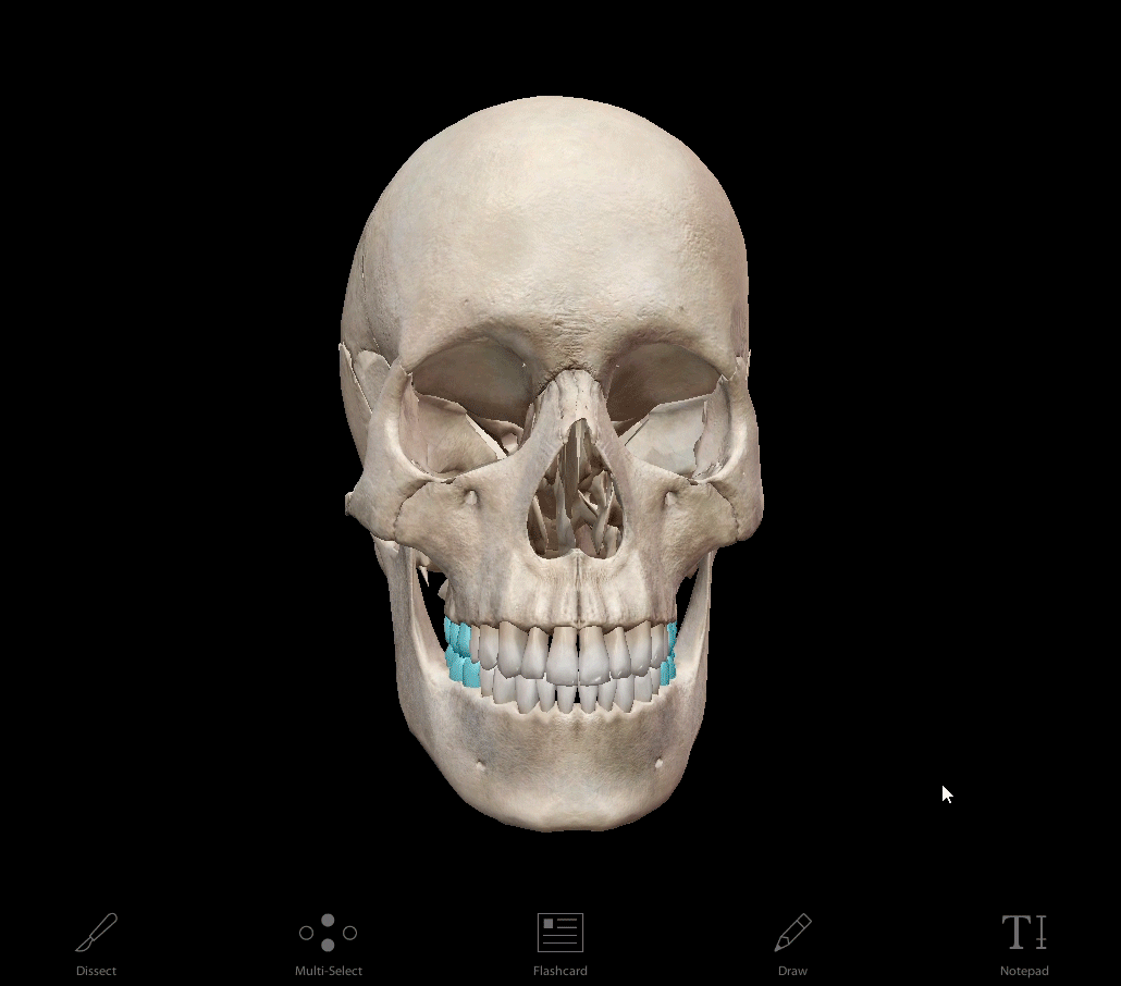
Canines
Canines are used for gripping and tearing food. They are also anterior teeth.
There are four canine teeth, one on each side of the incisors on both sides of the dental arch.
.jpg?width=515&height=386&name=screenshot%20-%202023-10-02T095142.282%20(1).jpg)
Image from Visible Body Suite.
Compared to the incisors, canine teeth are stronger and larger. The crown projects beyond the levels of the other teeth, and the upper canines (also called the eyeteeth) are larger and longer than their lower counterparts.
Premolars
There are eight premolars, also known as bicuspids. The bicuspid name comes from their shape: their crowns have two cusps separated by a groove. The upper premolars are bigger than the lower ones, though they are all smaller than the canines.
.jpg?width=515&height=386&name=screenshot%20-%202023-10-02T095336.401%20(1).jpg)
Image from Visible Body Suite.
Fun fact: baby teeth (aka deciduous teeth) do not include premolars!
Molars
Molars are used for chewing. Molars’ crowns are broad and flat, and of all the teeth, they have the largest surface for chewing.
GIF from Visible Body Suite.
There are twelve molars, which come in pairs of first, second, and third maxillary molars and first, second, and third mandibular molars.
The first upper molars have five cusps and three roots, while the second and third molars have three cusps and three roots. The first lower molars have five cusps, and the second and third lower molars have four or five cusps.
The third molars are known as the wisdom teeth, and they often don’t erupt. Their roots are more or less fused together. We said before that there are twelve molars, but some people have an odd number of third molars or none at all!
Learn more
You can read more about dental anatomy on the Visible Body Blog!
- Research Roundup: Why Good Oral Hygiene is Good for More Than Just Your Teeth
- 3 Dental Facts That Won’t Set Your Teeth on Edge
- Wise Up! Three "Whys" of Wisdom Teeth
Visible Body Suite and Courseware users can access free, interactive study content that will help you learn about or teach dental anatomy and surrounding structures—check out our free libraries of premade Flashcard Decks and Tours!
Be sure to subscribe to the Visible Body Blog for more anatomy awesomeness!
Are you an instructor? We have award-winning 3D products and resources for your anatomy and physiology course! Learn more here.

