New Anatomy Content: Fascia, Fascial Compartments, and Compartment Syndrome
Guess what, VB Blog readers? We have some exciting news for you! Fascial compartments have been one of our most-requested anatomy additions, and we've added them to Human Anatomy Atlas 2021 and Human Anatomy Atlas for Web Suite and Courseware!
To get you hyped for this new content, we’ll talk in this blog post about what fascia and fascial compartments are, and we’ll also give a brief overview of some common pathologies that affect them.
What is fascia?
 Fascia of the right arm. Image from Human Anatomy Atlas.
At the most basic level, fascia is a type of connective tissue. You might already be familiar with a few important instances of fascia in the body such as the iliotibial (IT) band, which runs along the outside of the thigh and stabilizes the knee, or the plantar fascia, which runs along the bottom of the foot and connects the heel bone to the toes.
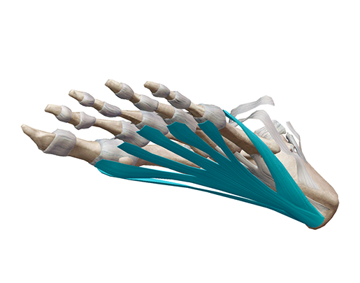
Plantar fascia. Image from Human Anatomy Atlas.
Fascia might not get as much publicity as ligaments or tendons, but it’s just as important. Thin layers of fascia surround our internal organs, as well as muscles, groups of muscles, blood vessels, and nerves, holding them in place or allowing them to slide past each other as needed.
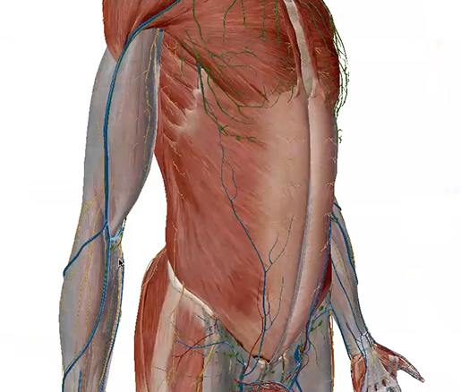
 Fascia of the thighs with nerves and blood vessels. Image from Human Anatomy Atlas.
In Human Anatomy Atlas, the fascia displays as part of the muscular system. For example, if you select the muscular system in the systems tray, the fascia will display along with all the muscles. In the image above, you can see the subcutaneous veins and nerves outside the fascia layer on the 3D model.
Like ligaments and tendons, fascia is composed primarily of elastin and collagen. This makes it flexible, so it can stretch when the body moves. When healthy, fascia is also usually smooth and slippery, able to shift its consistency between a more gel-like state and a more fluid one.
Here’s another cool fascia fact: fascia contains pain receptors and mechanoreceptors, which are sensory receptors that detect stretching or changes in pressure. For that reason, some scientists consider the body’s fascia to be one giant sensory organ!
What are fascial compartments?
Fascial compartments are what we call groups of muscles and nerves wrapped in a layer of fascia. Usually, muscles within the same compartment are innervated by the same nerve or nerves. Compartments are separated from one another by special bands of fascia called intermuscular septa.
There are four fascial compartments in each of the upper limbs: the anterior and posterior compartments of the arm, and the anterior and posterior compartments of the forearm.
The anterior compartment of the arm contains the coracobrachialis and the prime movers of elbow flexion, the muscles that flex the elbow joint: the biceps brachii and brachialis.
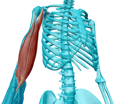 The muscles of the anterior compartment of the upper arm. Image from Human Anatomy Atlas.
In contrast, the posterior compartment of the arm contains the muscles that extend the elbow joint: the triceps brachii and anconeus. The anterior compartment of the forearm contains muscles that flex the wrist and digits and pronate the forearm, whereas the posterior component of the forearm contains muscles that extend the wrists and digits and supinate the forearm.
The thigh, or upper leg, has three compartments: anterior, medial, and posterior.
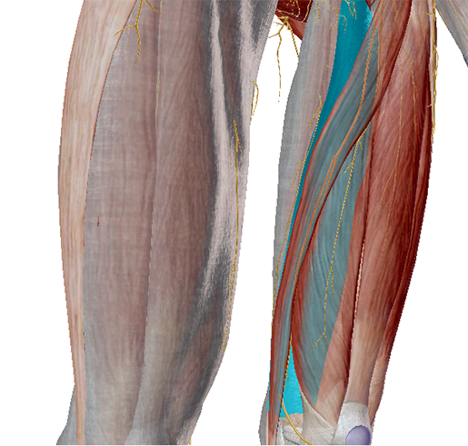 The medial intermuscular septum (highlighted) of the left leg, separating the anterior and medial compartments. Image from Human Anatomy Atlas.
The anterior compartment contains the sartorius, articularis genu, and the four muscles that make up the quadriceps femoris group. These muscles are primarily involved in the extension of the knee joint.
The medial compartment contains the pectineus, external obturator, gracilis, adductor magnus, adductor longus, and adductor brevis. Most of these muscles participate in the adduction of the hip.
The hamstrings (biceps femoris, semimembranosus, and semitendinosus) make up the posterior compartment. These muscles help the knee joint flex.
The lower leg has four compartments: anterior, lateral, deep posterior, and superficial posterior. The tibialis anterior, extensor hallucis longus, and extensor digitorum longus, and peroneus tertius, can be found in the anterior compartment. These muscles participate in the dorsiflexion of the foot.
The lateral compartment contains the fibularis brevis and fibularis longus. The deep posterior compartment contains the tibialis posterior, flexor hallucis longus, flexor digitorum longus, and popliteus. The popliteus helps rotate and flex the knee. The rest of the muscles in the deep posterior compartment participate in the flexion of the toes as well as the inversion and plantarflexion of the foot.
Lastly, the superficial posterior compartment houses the gastrocnemius, soleus, and plantaris. The gastrocnemius is a prime mover of knee flexion, and the plantaris and soleus participate in plantarflexion of the foot.
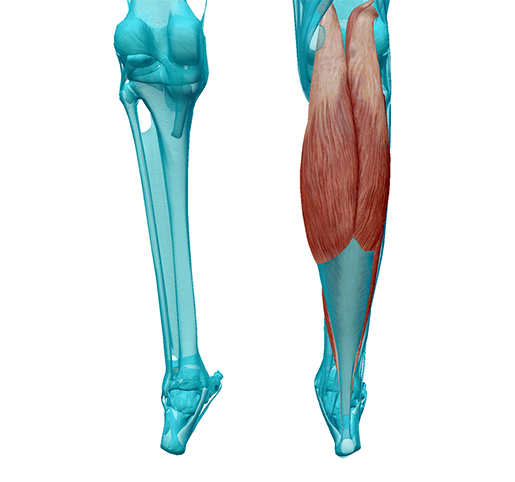 The muscles that make up the superficial posterior compartment of the lower leg. Image from Human Anatomy Atlas.
Injury to fascia
Fascia can become injured, much like tendons and ligaments. Plantar fasciitis, for example, happens when tension and stress on the plantar fascia cause tears to form in it, leading to inflammation and sharp pain.
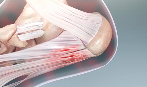
Microtears in the plantar fascia near where it attaches to the calcaneus bone. Image from Muscles & Kinesiology.
The IT band can become inflamed, too, when it’s overly tight and rubs against bone. These types of sports injuries reinforce the importance of balancing strength and flexibility, maintaining proper form when exercising, warming up and cooling down properly, and getting enough rest between exercise sessions.
Traumatic injury, surgery, overuse, or too little physical activity can lead to another type of problem with fascia—adhesions. When the texture of the fascia becomes more dry and sticky, it can tighten around muscles, causing pain and limiting mobility.
Regular exercise, stretching, and maintaining good posture are all good ways to help reduce the risk of injury to fascia.
Compartment syndrome
Compartment syndrome is a pathology specific to fascial compartments, and it can be either acute or chronic. In general, compartment syndrome happens when there is bleeding or swelling inside a compartment. This puts pressure on the fascia surrounding the compartment, as well as the nerves and blood vessels inside it.
Acute compartment syndrome usually results from a severe injury, such as a broken bone or crush injury. It can also occur after surgical repair of a blood vessel or as a result of a tight cast or bandage. Intense pain is the primary symptom, but a tingling or burning sensation and a feeling that an affected muscle is “tight” are also common. Acute compartment syndrome is an emergency medical condition, and needs to be treated as soon as possible. Prolonged pressure within a compartment can cause tissue death and damage to nerves. To relieve the pressure, medical professionals perform a procedure called a fasciotomy, in which they make an incision in the skin and fascia of the affected compartment.
Chronic compartment syndrome is usually brought on by exercise and relieved with rest. Cramping pain, swelling of the affected muscle, and tingling in the area around that muscle are common symptoms.
Want to learn more about connective tissue in the human body? Check out these VB blog posts:

Be sure to subscribe to the Visible Body Blog for more anatomy awesomeness!
Are you an instructor? We have award-winning 3D products and resources for your anatomy and physiology course! Learn more here.

