
3D Skeletal System: 5 Awesome Ligaments
How much do you enjoy moving? A lot, I bet. While many attribute our ability to move to the muscular system, it actually goes a little deeper than that! Your skeletal system is a rigid organ (yep, it’s living tissue!) that, without your awesome ligaments, wouldn't be able to move normally!
Ligaments are fibrous swathes of connective tissue that connect bones and help prevent your joints from flapping around willy-nilly. They also help to hold organs in place.
Let’s take a look at five of the coolest ligaments in your body!
5. Linea alba
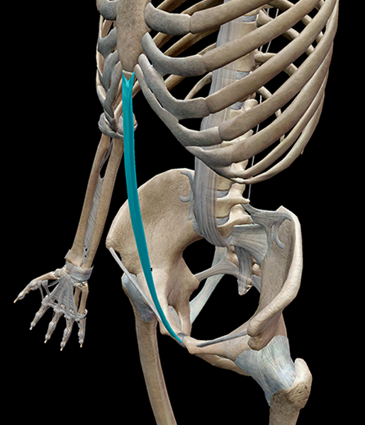 Image from Human Anatomy Atlas.
I love the linea alba. Do you know why? Because it’s exactly what its namesake says it is—a white line. The linea alba is a thin stretch of connective tissue that runs between the xiphoid process of the sternum and the pubic symphysis of the pelvic girdle. It also acts to divide the two rectus abdominis muscles.
4. Pubocervical fascia
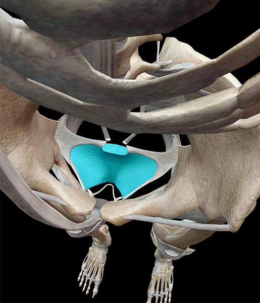
Image from Human Anatomy Atlas.
Stretched across the pelvic cavity is the pubocervical fascia, which connect the cervix to the pelvic walls and posteriorly blend with the perineal membrane. Think of the pubocervical fascia as a sort of hammock for the uterus and bladder.
3. Flexor retinaculum
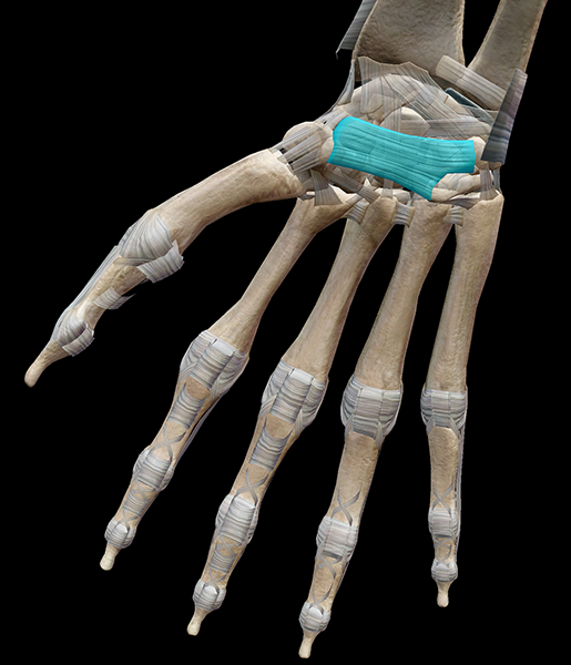
Image from Human Anatomy Atlas.
The flexor retinaculum and I have a complicated relationship. On the one hand (ha!), it helps to keep the flexor tendons together; on the other, it’s to blame for my increasingly rough ride through the carpal tunnel.
The flexor retinaculum is an arch of tough, fibrous tissue attached to the pisiform, hamate, trapezium, and scaphoid, and—with the carpal bones—forms a tunnel through which the flexor tendons of the hand and median nerve pass. Carpal tunnel syndrome occurs when one of the tendons becomes inflamed; since the retinaculum is so tough, there’s not enough stretch in it to accommodate the swollen tendon, so the tendon presses against the median nerve, resulting in numbness and/or pain.
2. Nuchal ligament
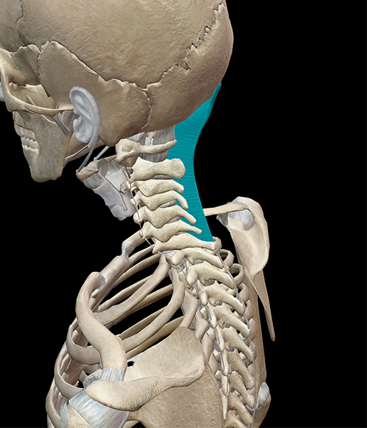
Image from Human Anatomy Atlas.
You know what’s awesome about the nuchal ligament? If you look at it laterally, it looks like a shark fin (yes, I’m one of those people who watches “Shark Week” religiously).
The nuchal ligament extends from the external occipital protuberance and median nuchal line to the spinous process of the seventh cervical vertebra. It stabilizes the head and neck, as muscles that would otherwise attach to the spinous processes of the vertebrae instead attach to the nuchal ligament.
1. All the ligaments of the skull
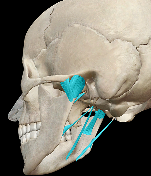
Image from Human Anatomy Atlas.
How could I pick just one ligament in the skull when all of them are so important? The ligaments of the skull comprise the ligaments that surround the temporomandibular joint, reinforcing the area where the cranium articulates with the mandible. The temporomandibular joint is a synovial joint and allows not only for flexion and extension, but also small movements of rotation and gliding.
The main “hinge” of the joint is the sphenomandibular ligament—a flat, thin band that connects the spina angularis of the sphenoid to the lingual of the mandibular foramen. The other ligaments connect the mandible to various bones of the skull.
| Did you know? Ligaments support multiple types of joints, including synovial joints and syndesmoses. |
Be sure to subscribe to the Visible Body Blog for more anatomy awesomeness!
Are you an instructor? We have award-winning 3D products and resources for your anatomy and physiology course! Learn more here.
Related Posts:
- 3D Skeletal System: Atlas, Axis, and the Atlanto-Axial Relationship

