Different Anatomy Tools Are Key to the Learning Process
We love to keep up to date with anatomy education research, so today on the Visible Body Blog, we’re going to look at a recent study on anatomy visualization. Published in BMC Medical Education, the study examines how students see the role of 3D images in anatomy education.
The authors are researchers from Karolinska Institute’s Department of Learning, Informatics, Management and Ethics. Karolinska Institute is one of the world’s best medical schools, and in addition to producing the most academic medical research in Sweden, it’s also home to the Nobel Assembly at the Karolinska Institute, which selects the Nobel Prize winners in Physiology or Medicine.
Through the university library, all students at the Karolinska Institute have access to Courseware, Visible Body’s learning and teaching platform that comes with a full suite of apps that feature over 6,000 3D, interactive structures.
3D images and models help students develop factual and spatial knowledge, and students view these tools as being effective and engaging, but as the authors point out, instructors need to understand the student perspective in order to design activities that will best help their students. Since experts like anatomy instructors look at 3D images differently than a less-experienced student, it’s important to know how students interpret these learning tools.
“To further understand how to facilitate and support the learning of anatomy, there is a need to know more about the student perspectives on how they can use and benefit from 3D images,” the authors write.
They set out to study how students interact with and use anatomy visualizations, focusing on images from real human bodies displayed digitally on a large visualization table.
Twenty-four students and one tutor participated in an anatomy training unit for the study.
The students were enrolled in medicine, physiotherapy, and nursing programs, and the tutor was an experienced medical student.
To see how students approached identifying anatomical structures, the researchers organized educational sessions where students explored the images and visualization table. The sessions emphasized clinical applications and self-direction as well as working in pairs.
The students looked at written clinical cases and then explored the images on the visualization table. The tutor was present to support the students.
The researchers recorded video and audio of the students doing this and also interviewed them and the tutor afterwards. Students found the images particularly interesting because they were scans from real people, and they liked how the images represented the variations between individuals not represented in textbooks. They enjoyed comparing images and their ability to zoom through different layers within the body to see spatial relationships.
They also found that the images were indistinct and difficult to parse.
“The students expressed a need to be able to refer to basic anatomical knowledge to make use of the 3D images. Such references could be to their prior anatomy studies, but also the simultaneous use of other learning resources, such as an atlas or internet-based learning resources,” the authors write.
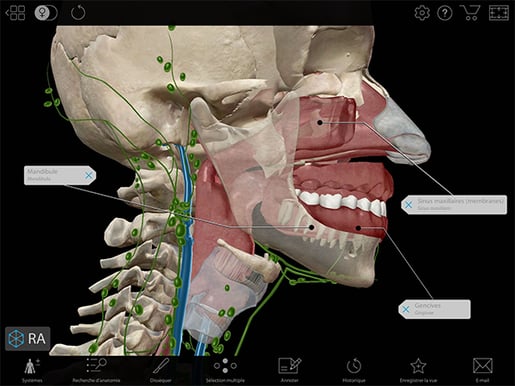 Image from Human Anatomy Atlas 2022 +.
Students said that other tools where the structures are clear and named would be more appropriate for certain stages of learning. One student said, “I’m thinking of Visible Body for instance. It’s excellent in that way. You can just click and it recognizes every vessel and then you get the whole vascular system.”
Students valued the ability to explore the 3D models on their own because it allowed them to focus on what they needed. The tutor said that because of this, they covered less material than usual, but they had a greater depth of understanding.
The students and tutor “suggested that 3D images in applications such as Visible Body may be helpful since anatomical structures are clearly distinguished by colours compared to CT or MR images of real bodies.”
 GIF from Human Anatomy Atlas 2022 +.
Visible Body helped them get a baseline knowledge they could then apply to the complex, layered, more difficult real images.
The findings suggested that using a combination of different learning resources will help students learn best.
Read more
Want to learn more about Visible Body and 3D learning? Check out these links!
- Visible Body Improves Student Performance: HAPS 2021 with Dr. Cindy Harley
- Can Augmented Reality Improve the Learning Experience of Future Healthcare Professionals? (2022)
- Is Visible Body Courseware a Good Alternative to an Anatomage Table?
Be sure to subscribe to the Visible Body Blog for more anatomy awesomeness!
Are you an instructor? We have award-winning 3D products and resources for your anatomy and physiology course! Learn more here.

