3D Urinary System Lesson Plan with Visible Body
 An assignment in Courseware.
Lecture in 3D
Visible Body has created the ultimate lecture aid: Tours. A Tour is like if a PowerPoint presentation was made up of interactive 3D models rather than boring 2D slides. We’ve even got a library of premade Tours that are free for all users!

GIF from VB Suite.
When it comes to lecture, a good place to start is the premade kidney Tour. This Tour takes you through kidney anatomy, from the location of the kidneys to the microanatomy model showing a cross-section.
Want to make your own Tour? It’s easy to customize Views and assemble them into a Tour.
- Use VB Suite’s search and filter tools to find exactly what you want. From VB Suite’s main menu, click Human Anatomy and enter your query into the “Filter results by name” box above all the Views. Click on the filter icon to select the kind of content you’re looking for, like a gross or microanatomy model, animation, or histology slide.
- Once you’ve selected your first view, it’s time to get creative. Using your mouse or keyboard, position the model exactly how you’d like. You’ll be able to manipulate the model as you present it to your students, so unlike a traditional slideshow, you’re not stuck with a static image. Using the Draw tool, you can add 3D annotations like arrows, shapes, and freehand drawings. The Notepad allows you to add text to the View, and you can also add tags.
- Click “Save View” and give your custom View a name!
- Once you’ve created several Views, you can combine them into a Tour. Navigate back to the main screen and open the “Tours” section. Click "Start" and select the Views you saved in the order you want them to appear. You can then save the Tour.
You can share a Tour with a few easy clicks—you can use a share link, or you can generate a QR code for your students.
Below is an example of how you can teach the male and female urinary systems with a custom Tour!
Open the Tour and use a view of renal vasculature to show how blood enters the kidney through the renal arteries, where they then branch into many capillaries inside the kidney. You can zoom in and rotate the model as needed to give students a more dynamic look at the structures.
Then you can select the arrow to move to the next View and use the nephron microanatomy model to explain the ~1 million nephrons in each of the kidneys. Each one filters waste out of the blood and produces urine. Zoom in and show your students the glomerulus to illustrate where filtration begins. Highlight the convoluted tubules where filtrate passes through.
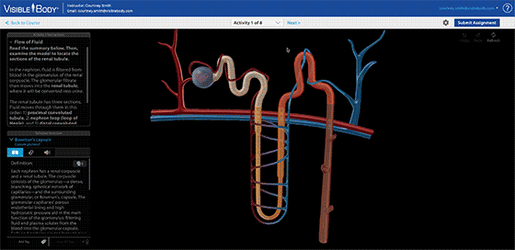
Use a View of the simplified nephron model to show the flow of filtrate from the glomerular capsule to the renal tubule. As fluids flow through the renal tubules, vital substances are reabsorbed into adjacent capillaries. Concurrently, waste products pass from the capillaries into the renal tubules and move as urine into the collecting duct.
Now, return to the first gross anatomy View and show the path of urine out of the kidney as it moves through the renal pelvis, down the ureter into the bladder, and finally out of the body through the urethra.
Make review easy
If you’ve covered all the parts of the kidney with your students, click on the Study tab at the top of the menu and scroll down to find urinary system quizzes. You can ask different students to come up and select the correct structure.
 GIF from VB Suite.
Flashcards are another great review activity—you can make your own Flashcards, or you can choose from Visible Body’s free library of Decks. You can use Flashcard Decks on your classroom’s projector to get the entire class involved, or you can even have students make their own Decks for an assignment.
Visible Body Customer Engagement Specialist and real-life A&P instructor Jenn Smulligan designed this great Flashcard activity for her students. That particular assignment is geared towards bony landmarks, but it can easily be adapted for other anatomy! Check out that blog post for a breakdown of the assignment and a copy you can use and customize in your Courseware course.
Lab activities in Courseware
Using Visible Body Courseware, you can assign interactive, engaging assignments that break big concepts down into bite-sized pieces.
Courseware assignments create an “on the rails,” distraction-free online environment where students can move seamlessly through activities. Courseware assignments combine 3D gross anatomy and microanatomy views, videos, illustrations, and histology slides interspersed with short quizzes to check comprehension
 An assignment in Courseware.
The Visible Body team has created premade assignments that cover all major body systems and introductory biology units. Instructors can customize those assignments, or they can build their own from scratch.
Courseware has two premade lab activity assignments regarding the urinary system. To illustrate how these assignments walk students through difficult concepts, let’s take a closer look at the premade Nephrons and Glomerular Filtration lab activity.
The assignment starts by walking students through a 3D nephron model to introduce the renal corpuscle and glomerular filtration, followed by a urine filtration video that shows students how urine is filtered through the glomerular capillaries. Next, a histology slide shows students more about glomerular structure and the location of the podocytes. A quick, four-question quiz checks students’ understanding of these concepts, and then it’s time to deepen understanding with an illustration and a histology slide that show structures that affect the glomerular filtration rate. Students then return to the nephron model to learn about fluid pressure and the arterioles. The assignment ends with another quick quiz.
Students' quiz grades and time-on-task automatically report to the Gradebook. Courseware connects with many LMS platforms to make course integration seamless. You can learn more about assignments here!
We hope this blog post has given you ideas for your next urinary system lesson plan! If you're looking for more, check out this library of premade courses for Courseware and this awesome active learning workshop webinar on the renal system delivered by Dr. Cindy Harley.
Want to try Courseware for yourself? You can sign up for a free instructor trial.
Be sure to subscribe to the Visible Body Blog for more anatomy awesomeness!
Are you an instructor? We have award-winning 3D products and resources for your anatomy and physiology course! Learn more here.

