

Behind the Scenes of Visible Biology: Bringing the Blood Cell Lab into 3D and AR
[Update: All of Visible Body's biology content is exclusively available in VB Suite!]
Recently, Visible Body partnered with Google’s search team to create over 15 new 3D and AR models illustrating important biology concepts and structures, including prokaryotic and eukaryotic cells and chromosomes, photosynthesis, monocot and dicot plant structure, and the different types of blood cells. The editorial team created (free!) lab activities to accompany these interactive 3D models, allowing instructors to teach engaging lessons online.
Bridging the gap between what you can do in an in-person lab versus an online one is a unique challenge—one that the editorial and content teams at Visible Body addressed carefully when creating the models and activities for the 3D blood cells lab.
Today, we’re going to talk about how Visible Body’s 3D blood cell models differ from blood smear microscope slides, what they offer that slides don't, and how students can use the 3D models and histology slides together to learn about blood cells in the Introduction to Biology course in Visible Body Courseware.
What does a blood smear on a microscope slide look like?
Creating peripheral blood smears is the typical method researchers use to study the histology of blood. Pathologists also use blood smears to count the different types of blood cells and analyze their size, shape, color, spatial distribution, and relative abundance. This is useful in the diagnosis of blood disorders such as anemia, sickle cell anemia, and leukemia.
In a peripheral blood smear, a drop of peripheral blood (the blood that circulates throughout the body) is spread into a thin, single layer of blood cells on a microscope slide. You can apply different types of stains to the slide to highlight different types of blood cells—some blood cells and their components show up better with an acidic stain, and others are better viewed with a basic stain. Hematoxylin and eosin (H&E) staining is commonly used in histology slides.
Histology slides are commonly used in biology and A&P lab courses to show the visual differences between different types of blood cells. For example, it’s easy to see that red blood cells and platelets don’t have nuclei, whereas white blood cells have nuclei—many of them large and multi-lobed. The granules of certain white blood cells, known as granulocytes or granular myeloid white blood cells, are also visible on histology slides.
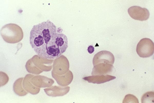
This photomicrograph of a fixed blood smear shows a granular myeloid white blood cell (a polymorphonuclear leukocyte), red blood cells, and a cluster of platelets (identified with an arrow). Image from the CDC’s Public Health Image Library.
How do Visible Body's 3D blood cell models differ from what you'd see in a blood smear?
When you look at a blood smear using a microscope, you’ll see a single layer of blood cells that look flat and often overlap each other, making it difficult to see the shape and structure of each cell.
In contrast, when you look at the 3D models in the blood cells unit on the Biology Learn Site, you’ll notice that they are designed to look like droplets of blood before they are smeared but with histological stains already applied. This allows you to observe the shape, size, structure, and stained color of various types of blood cells suspended in plasma.
![]() Here you can see the red blood cells and platelets suspended in plasma. Image from the Biology Learn Site.
Here you can see the red blood cells and platelets suspended in plasma. Image from the Biology Learn Site.
In addition, there are a few important structural characteristics of blood cells that aren’t visible using a normal light microscope that are included in Visible Body’s 3D models. For example, the 3D model of lymphoid white blood cells shows microvilli, which you can’t see on a typical histology slide.
![]() Lymphoid white blood cells with microvilli. Image from the Biology Learn Site.
Lymphoid white blood cells with microvilli. Image from the Biology Learn Site.
Lastly, while a blood smear can contain various types of blood cells in different proportions, each of the 3D models was created to feature a specific group of blood cells for comparison purposes.
Red blood cells are present in all the models because they are the most numerous type of blood cell. The red blood cell and platelet model shows both inactivated and activated platelets to demonstrate the structural differences between them. Similarly, the agranular myeloid white blood cell model contrasts an inactivated monocyte with an activated monocyte, complete with pseudopodia.
![]() An activated vs. an inactivated monocyte in a blood droplet.Image from the Biology Learn Site.
An activated vs. an inactivated monocyte in a blood droplet.Image from the Biology Learn Site.
The granular myeloid white blood cell model shows the size of the granules and the size and shape of the nucleus in a neutrophil, basophil, and eosinophil. The lymphoid white blood cell model uses color and the presence of granules to differentiate between B and T cells (agranular) and natural killer cells (granular).
What was it like to work on the 3D models and labs?
Adrienne Devlin, Executive Editor at Visible Body, is one of the team members who worked on the blood cell labs and models. Here’s what she had to say about her experience working on the project:
“Out of all the biology labs and models we created, the blood cell ones were the most challenging and rewarding. When I started researching blood cell lab activities and talking to instructors, a few things became clear to me: 1. Studying histology slides is a huge part of the lab, 2. Students struggle to distinguish between the different blood cell types in histology slides, and 3. No one had ever created an interactive, 3D representation of a histology slide.
Based on my research, I created the blood cell lab activities with a few key goals. I grouped the blood cells into commonly used categories to make it easier for students to compare their structure and functions. I created pre-lab activities that introduce students to using histology images and lab activities that allow students to explore interactive 3D models to identify the different types of blood cells. I worked closely with the artists and instructors to create our blood cell models in a way that balances accuracy and usefulness in a biology lab course with the artistic license needed to make the blood cells 3D, interactive, and comparative.
The most rewarding part of the process was showing the lab activities and 3D models to instructors and hearing how excited they are to use this content to teach their students about blood cells.”
Hillary, a 3D Medical Artist at Visible Body, worked on the concept sketches for the models and the granular myeloid and agranular lymphoid models. She said that the biggest challenge of the project was that “it was tough dealing with the technical constraints the models had to fit into while also creating an engaging, unique tool that still effectively teaches the concepts we wanted the audience to understand.” But the final product was worth it! Hillary commented that “it was cool to see that work out in the end.”
Anne, another VB 3D Medical Artist who worked on the blood cell models, said that “Working on a project that starts out as an idea in someone's brain and turns into a functional (not to mention beautiful!) teaching tool is an awesome experience for a medical animator.” The biggest challenge she found in working on the project was “representing various textures and levels of transparency to achieve what was a believable drop of blood plasma full of microscopic cells.” She was also pleased with the final product, saying that “this is a great team to get to be a part of and we are happy with what we were able to accomplish together!”
How can students use 3D models to study Blood Cells?
When creating the blood cell models for the Biology Learn Site and lab activities, the Visible Body content team combined the information you’d observe from viewing a peripheral blood smear under a microscope with the ability to explore structures that’s only really possible by interacting with a 3D model.
Using Visible Body’s free Biology Learn Site, students can read through brief articles describing the essential information about each type of blood cell. Each article contains one of the four 3D blood cell models: red blood cells and platelets, granular myeloid white blood cells, agranular myeloid white blood cells, and lymphoid white blood cells. Students can rotate and zoom into the models to visualize and compare the shapes and sizes of the different types of blood cells. Students can get a more traditional blood smear-like view by rotating the models to look at them from above (as in the image below).
![]() Top-down view of the granular myeloid white blood cells model. Image from the Biology Learn Site.
Top-down view of the granular myeloid white blood cells model. Image from the Biology Learn Site.
On compatible devices, students can even bring the blood cell models into their environment using augmented reality.
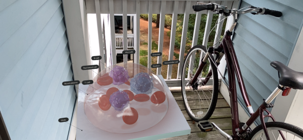 The lymphoid white blood cells model in the great outdoors!
The free lab activities on the Biology Learn Site offer lab activities corresponding with each Learn Site article and 3D model. Students can use the Blood Cells Glossary, which contains all the 3D blood cell models and definitions for their labels, as an additional study tool.
Use Courseware for a side-by-side comparison between 3D models and histology slides.
Along with the Biology Learn Site, Visible Body offers a premade course for Courseware, which includes versions of the blood cell lab activities that feature histology slides for each type of blood cell from Visible Body’s A&P app.
With the Courseware versions of the blood cell lab activities, students can get the best of both worlds—real histology slides from the A&P app and interactive 3D and AR models from the Learn Site—without the need for a microscope or a physical lab space. If you have a Courseware instructor account, you can download the Introduction to Biology course from our premade courses page.
Be sure to subscribe to the Visible Body Blog for more anatomy awesomeness!
Are you an instructor? We have award-winning 3D products and resources for your anatomy and physiology course! Learn more here.
