Teaching Cell Types: Differentiated Instruction with Visible Body
Prokaryotic versus eukaryotic? Plant versus animal? Learning the distinctions between cell types is an important step in learning about function and thinking critically about structures within the cell. Visible Body Suite has 3D models that bring the microscopic into full view, giving students a way to visualize and interact with tiny structures.
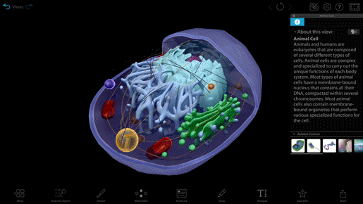
Animal cell model in VB Suite.
Today on the blog, we’ll look at teaching cell types through the lens of differentiated instruction. Differentiated instruction is the concept that every student is different, so a one-size-fits-all approach to instruction won’t fit. There are three areas of instruction that can be differentiated: content, process, and product.
We’ve got a whole blog post on differentiated instruction and Visible Body, but let’s look at how you can use differentiated instruction to teach the differences and similarities between cell types!

Content
With differentiated content, instructors give students multiple ways to receive information. VB Suite provides many different avenues for learning in order to maintain student engagement.
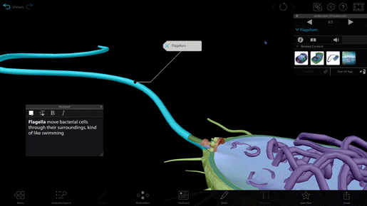
Tour made in VB Suite.
Make lectures interactive
Tours are a series of saved views—imagine a presentation made up of interactive 3D slides. With Tours, you can make your visual aids come alive. VB Suite contains full 3D models of a plant cell, an animal cell, and a bacterial cell. The plant and animal cells are dissectible, giving you and your students the ability to closely examine organelles.
Provide your students with a share link to access the Tour at home and explore the models on their own. VB Suite’s built-in QR code function makes custom content super easy to share!
Read and listen
Want to know exactly what a cell wall is? Click the book icon in the info box! Each structure and view in VB Suite features an easily accessible, textbook-level definition. Students can click on the megaphone icon to hear how the structure's name is pronounced.
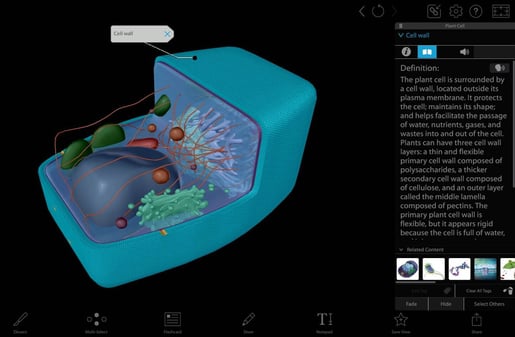
Plant cell model in VB Suite.
The info box also contains the Listen button—click on the talking head icon and your computer or mobile device’s text-to-speech tool will read the definition aloud. Multitaskers can listen to the definition as they use the Draw tool or Notepad to take notes!
Process
Once students have a foundational understanding of the difference between prokaryotic and eukaryotic cells and between plant and animal cells, it’s time to reinforce that knowledge. Instructors can differentiate this process by offering options for how students can, well, process the information they’ve just learned.
Find and identify organelles
Students can manipulate 3D models to find and identify organelles unique to the different cell types.
You can find structure identification activities as part of the premade prokaryotic and eukaryotic cells lab! Check out Visible Body’s treasure trove of lab manuals here. 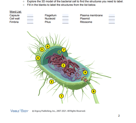
Screenshot of a lab activity in VB Suite.
Jigsaw
Break up students into groups. Each student in a group is responsible for becoming the group “expert” on a particular type of cell—assign a model to each student. After students work independently to gather information, the members of each jigsaw group puts what they’ve learned together.
Graphic organizers
A graphic organizer like a chart is a simple way to show the differences and similarities between cells.
Here's a hint—the premade lab manual we mentioned earlier features an activity that uses a table to compare animal and plant cells!
Product
It’s finally time for students to show what they’ve learned! For some, that means taking a test, but through differentiating product, instructors can offer alternative means of assessment.
Presentation
For some students, it’s more intuitive to talk through ideas rather than writing them down. Using 3D models as a visual aid, ask students to present the main differences and similarities between prokaryotic and eukaryotic cells and between plant and animal cells. Students can even make their own Tours, and this option lends itself well to group work. Students can record their presentations and submit the videos online—make sure to add a time limit to keep grading manageable!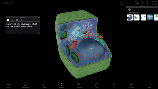
Plant cell model in a lab activity for in VB Suite.
Drawing
Using the 3D models and definitions for reference, more artistically inclined students can make a series of visual Venn diagrams, drawing and coloring the organelles rather than simply jotting down names. Students should label their drawings and provide 2-3 brief bullet points describing each organelle’s function.
DIY Flashcard Deck
Students can create their own Flashcard Decks to try to stump their peers!
Students should write a prompt on the front side (e.g., “This organelle contains digestive enzymes enclosed in a membrane.”). On the back, students should use the auto-tag function to identify the organelle and add text to identify the type of cell.
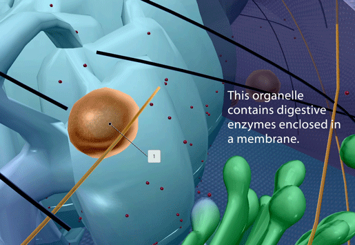
Flashcard in VB Suite.
Wrap-up
Differentiated instruction and VB Suite are a great fit—VB Suite has the flexibility and multimodality to get many different learners engaged.
Do you use VB Suite as part of differentiated instruction? Drop us a comment below!
NGSS Standards
The above lesson plan fits these Next Generation Science Standards (NGSS):
HS-LS1 From Molecules to Organisms: Structures and Processes. Students who demonstrate understanding can:
- Construct an explanation based on evidence for how the structure of DNA determines the structure of proteins which carry out the essential functions of life through systems of specialized cells. (HS-LS1-1)
- Develop and use a model to illustrate the hierarchical organization of interacting systems that provide specific functions within multicellular organisms. (HS-LS1-2)
- Use a model to illustrate the role of cellular division (mitosis) and differentiation in producing and maintaining complex organisms. (HS-LS1-4)
- Construct and revise an explanation based on evidence for how carbon, hydrogen, and oxygen from sugar molecules may combine with other elements to form amino acids and/or other large carbon-based molecules. (HS-LS1-6)
HS-LS3 Heredity: Inheritance and Variation of Traits. Students who demonstrate understanding can:
- Ask questions to clarify relationships about the role of DNA and chromosomes in coding the instructions for characteristic traits passed from parents to offspring. (HS-LS3-1)
- Make and defend a claim based on evidence that inheritable genetic variations may result from: (1) new genetic combinations through meiosis, (2) viable errors occurring during replication, and/or (3) mutations caused by environmental factors. (HS-LS3-2)
Did you know that the Visible Body Education Team has created a library of lesson plans and lab activities? Here's where you can find these resources, complete with NGSS standards.
Be sure to subscribe to the Visible Body Blog for more anatomy awesomeness!
Are you an instructor? We have award-winning 3D products and resources for your anatomy and physiology course! Learn more here.

