Spine Time: A Guide to Spinal Anatomy
Want to learn about spinal anatomy? We’ve got your back. In this blog post, we’ll talk about the bones, discs, muscles, and nerves that make your spine the body’s main support system.
First, let’s talk about vertebrae! The spine has 33 vertebrae, small bones that stack on top of each other.
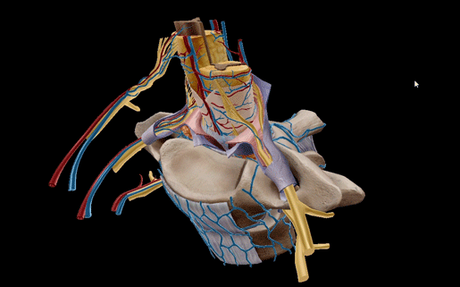
Cross section vertebrae model in Visible Body Suite.
The vertebrae are divided into five categories: cervical, thoracic, lumbar, sacral, and coccygeal.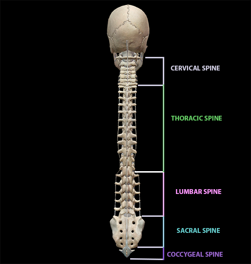
Image from Visible Body Suite.
Cervical vertebrae
The cervical vertebrae are the smallest vertebrae! They are located in the neck.
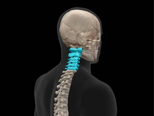
Image from Visible Body Suite.
The most superior vertebra is C01, the atlas. The atlas is set apart from the others because it’s ring-shaped and lacks a body and spinous process. The spinal cord passes through the middle of the ring.
The atlas works with the axis, aka C02, to support the skull and give a range of motion to the neck. A unique feature of the axis is the odontoid process, which articulates with the atlas to create a flexible pivot joint.
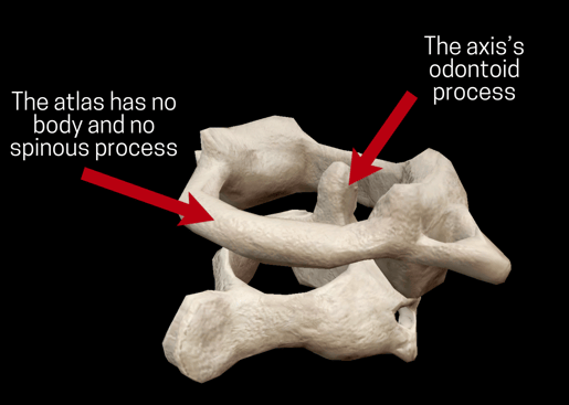
Image from Visible Body Suite.
Vertebrae C02-C07 consist of an anterior body and a posterior vertebral arch, and the spinal cord runs between those two parts in an opening called the vertebral foramen. The vertebral arch is made up of two pedicles and two laminae. The body bears weight and provides surfaces for the attachment of intervertebral discs.
In the case of C07, the most inferior cervical vertebra, the vertebral foramen is usually small or absent, and the vertebral artery and vein pass in front of the transverse process.
Thoracic vertebrae
The thoracic vertebrae are T01-T12, the eighth through nineteenth vertebrae in the spine. .jpg?width=515&height=386&name=screenshot%20-%202023-09-07T110737.959%20(1).jpg)
Image from Visible Body Suite.
With the exception of T11 and T12, the transverse processes articulate with the tubercules of vertebrosternal and false ribs, and facets on the lateral sides of the body articulate with the heads of the vertebrosternal and false ribs.
Like most of the cervical vertebrae, the thoracic vertebrae have vertebral foramens. An intervertebral foramen is formed where the body and arch of two vertebrae articulate, allowing for transmission of the spinal nerves.
Lumbar vertebrae
The 20th through 24th vertebrae are the lumbar vertebrae, L01-L05. 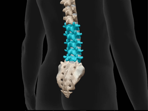
Image from Visible Body Suite.
In thoracic vertebrae, the transverse processes are located in front of the articular processes. The transverse processes’ superior tubercle connects with the superior articular process to create the mamillary process, and the inferior tubercle is called the accessory process.
At or around L03, the spinal cord ends and the cauda equina, a group of nerve roots that innervate the pelvis and lower limbs, begins. This is why lumbar punctures typically are performed between L04 and L05, to minimize any damage to the spinal cord.
The sacrum
Your spine doesn’t stop at the lumbar vertebrae! The 25th through 29th vertebrae, S01-S05, fuse in early adulthood to form the sacrum. The sacrum is a triangular bone located between the hip bones.
The shape of the sacrum varies depending on sex: males tend to have taller, thinner sacrums than females, whose sacrums allow for more space in the pelvic cavity.
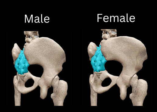
Note the difference in sacrum and pelvis shape. Images from Visible Body Suite.
The coccyx
Last but not least, the final four vertebrae fuse to make the coccyx, also known as the tailbone. The first vertebra in the coccyx resembles a lumbar vertebra, but the next vertebrae diminish in size until the last vertebra, which often is just a nodule.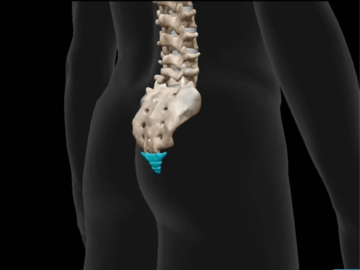
Image from Visible Body Suite.
The surfaces of the coccyx provide attachments for several ligaments, and the anterior surface of the coccyx supports part of the rectum. The coccyx’s terminus is attached to the tendon of the external anal sphincter.
Intervertebral discs
Like we’ve mentioned before, between the vertebrae are discs. Intervertebral discs serve as shock absorbers: the middle of the disc is soft and pulpy, and when pressure is applied, the center becomes flatter and pushes against the fibrous external layers. In addition to shock absorption, the intervertebral discs help support the spine. 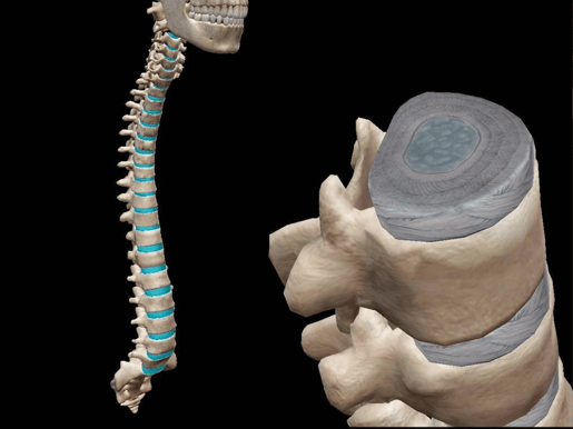
Image from Visible Body Suite.
The outer layers that encapsulate the center of the disc are called the annulus fibrosus. The outermost layer is made of fibrocartilage, which surrounds layers of cartilage fibers. The annulus fibrosis resists torsion, flexion, and extension.
Muscles
Given that the spine has 33 vertebrae, it makes sense that there are many muscles involved in the movement of the vertebral column! Since there are so many, in this blog post, we’re just going to cover the muscles involved in spine flexion, extension, and lateral flexion as well as thoracic spine rotation.
Spine flexion
Spine flexion is when you bend forward, rounding your back and moving your face and chest toward the ground, as if you’ve dropped your keys on the ground. The agonists, or prime movers, of spine flexion are:
- Rectus abdominis
- Internal oblique
- External oblique
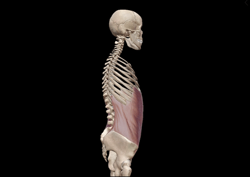
GIF from Visible Body Suite.
Spine extension
Spine extension is when you bend backward, arching the back, as if you’re looking up at a beautiful starry night. There are many agonists at play here:
- Spinalis cervicis
- Spinalis thoracis
- Longissimus capitis
- Longissimus cervicis
- Longissimus thoracis
- Iliocostalis cervicis
- Iliocostalis thoracis
- Iliocostalis lumborum
- Semispinalis capitis
- Semispinalis capitis
- Semispinalis cervicis
- Semispinalis thoracis
- Splenius capitis
- Splenius cervicis
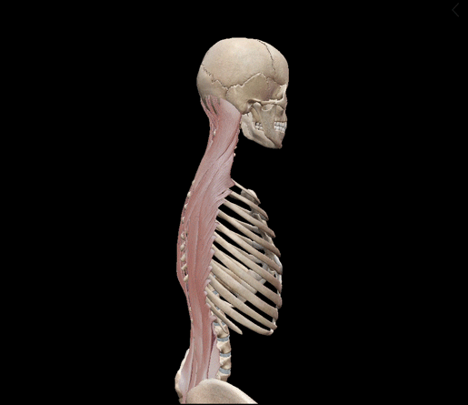
GIF from Visible Body Suite.
Spine lateral flexion
Spine lateral flexion is when you bend your spine to the side, moving your torso away from the center of your body, like when you’re leaning over to grab a water bottle in the door of a car. Spine lateral flexion agonists include:
- Spinalis cervicis
- Spinalis thoracis
- Longissimus capitis
- Longissimus cervicis
- Longissimus thoracis
- Iliocostalis cervicis
- Iliocostalis thoracis
- Iliocostalis lumborum
- Quadratus lumborum
- Latissimus dorsi
- Internal oblique
- External oblique
- Anterior scalene
- Middle scalene
- Posterior scalene
- Splenius capitis
- Splenius cervicis
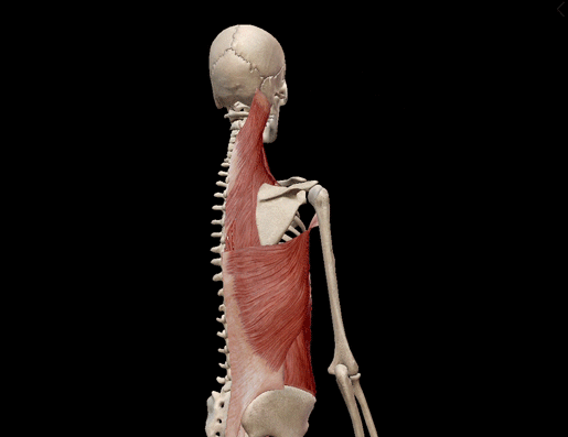
GIF from Visible Body Suite.
Thoracic spine rotation
Last but not least, let’s look at thoracic spine rotation! Thoracic spine rotation is when you twist the torso to either side, like you’re starting to turn around to glare at the person kicking the back of your seat on an airplane. The agonists at play here are:
- Internal oblique
- External oblique
- Rotatores breves
- Rotatores longi
- Multifidus
GIF from Visible Body Suite.
Nerves of the spine
The spine is home to the spinal cord—no duh! The spinal cord is one of the two major structures of the central nervous system, alongside the brain. It extends from the atlas down to the first or second lumbar vertebra, running through the spinal cavity. The spinal cavity is the term for the canal in the vertebral foramen.
There are 31 pairs of spinal nerves that branch out from the spinal cord and “peek out” from the neural foramina, the spaces between the vertebrae. These nerves continue to branch out, reaching all the corners of the body.
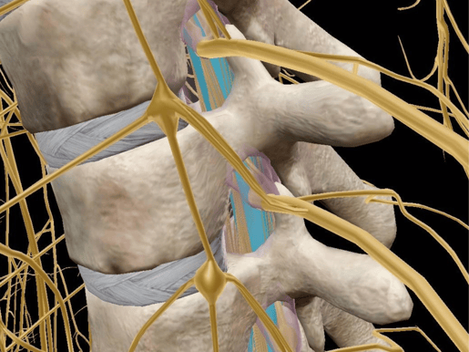
Spinal cord highlighted in blue. Image from Visible Body Suite.
Want to learn about spinal nerves in more detail? Check out this entire blog post we have on the topic!
Conclusion
The vertebrae are made up of five distinct sections: cervical, thoracic, lumbar, sacral, and coccygeal. Between these vertebrae are intervertebral discs, which act as cushion and support. The spine is very flexible, and many, many muscles help move it. The spine is also home to the spinal cord, a core part of the central nervous system from which most nerves branch.
Want to learn more about the spine? Check out these blog posts!
- When Your Spine Feels Less Than Fine: Common Spinal Pathologies
- What Is Scoliosis?
- In a Pinch: The Anatomy and Pathology of Cervical Radiculopathy
Visible Body Suite and Courseware users can access free, interactive study content on the spinal cord, vertebral column, muscles of the back and spine, and more—check out our free libraries of premade Flashcard Decks and Tours!
Be sure to subscribe to the Visible Body Blog for more anatomy awesomeness!
Are you an instructor? We have award-winning 3D products and resources for your anatomy and physiology course! Learn more here.

