

Investigating Renal Anatomy with Human Anatomy Atlas 2020
By now I'm sure you've heard the amazing news: Human Anatomy Atlas 2020 has made its debut! Along with the awesome pulmonary and thoracic structures Laura briefed you on a few weeks ago, we've got plenty more updates to brag about. Today I'm going to cover the newest features in the ol' renal department.
1. Vasculature Galore
Let's start off with circulation. In Atlas 2020 we've got TONS of new arteries and veins, but specifically I'm going to point you towards the renal ones. You can see how the renal arteries branch all the way down to the interlobular arteries, especially if we hide the renal columns.
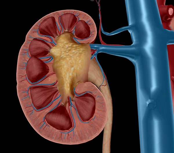 Image from Human Anatomy Atlas 2020.
The interlobular arteries are located between the renal lobules of the kidneys, but before reaching them, blood from the renal arteries flows through the segmental arteries, interlobar arteries, and the arcuate arteries. The interlobular arteries then branch outward from the arcuate arteries to supply the cortical tissue, which branches further into afferent arterioles supplying the nephrons.
2. Renal Sinuses
If you slice open the kidney you can clearly see we've amped up our texture and detail. The big yellow blob in the center is the renal sinus. If you cut him in half too, you'll see he's hollow! This baby is the kidney's central cavity, containing the renal pelvis, renal calyces, blood vessels, nerves, and adipose tissue (fat).
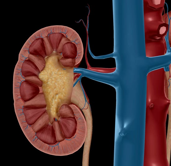 Image from Human Anatomy Atlas 2020.
3. Renal Calyces
Next, let's touch on the calyces, the pelvis's protrusions hidden inside the sinus. The pelvis itself is the funnel-shaped structure that joins up with the ureter. It functions as an excretory channel for the kidneys. But before the urine can get to the pelvis, it hits the calyces. At the apex of the renal pyramids, urine passes through a renal papilla into a minor calyx, which converges with some friends to form a major calyx.
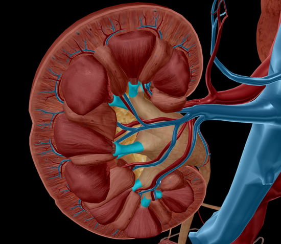 Image showing minor calyces from Human Anatomy Atlas 2020.
We only had these calyx guys selectable in our microanatomy model before, but for the 2020 release we went ahead and added them in the main model set for accuracy as well as to appease the masses.
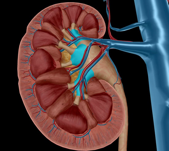 Image showing major calyces from Human Anatomy Atlas 2020.
4. Renal Columns
The renal medulla consists of numerous pyramids, which are striated conical masses of tubules. These pyramids have wide bases that face the renal cortex and narrow into apices, called papillae, as they converge towards the pelvis. The soft and granular renal cortex arches over the bases of the pyramids and dips in between them, forming renal columns. These columns are composed of blood vessels, urinary tubes, and fibrous material. All of these help to anchor the renal cortex, which with the medulla, contains the nephrons: the functional units of the kidney.
 Image from Human Anatomy Atlas 2020.
Both the pyramids and columns have gotten a new look as well. I personally like to refer to the 2020 versions as "plump," because in my opinion, it's more whimsical than just saying "realistic" or "anatomically correct."
We've also got a nifty YouTube video showing you basically everything I just went over (if you're not yet convinced you need to get your hands on Atlas 2020).
For more Human Anatomy Atlas 2020 updates, subscribe to the blog and subscribe to our YouTube channel. We've got tons of great things cooking!
Be sure to subscribe to the Visible Body Blog for more anatomy awesomeness!
Are you an instructor? We have award-winning 3D products and resources for your anatomy and physiology course! Learn more here.
