Exploring Thoracic and Pulmonary Structures with Human Anatomy Atlas 2020
We’ve talked about the thoracic cage and the respiratory system on the Visible Body blog before—having lungs is important, after all! With the release of Human Anatomy Atlas 2020, we’ve got some updates to thoracic and pulmonary structures that make them even more detailed, visually stunning, and fun to study! We’re super excited to share them with you.
1. Radiate sternocostal ligaments
First off, we’ve got the radiate sternocostal ligaments. These fibrous bands of tissue attach the sternal ends of the costal cartilages of ribs 1-7 to the sternum. They help give the ribs the mobility they need to accommodate breathing.
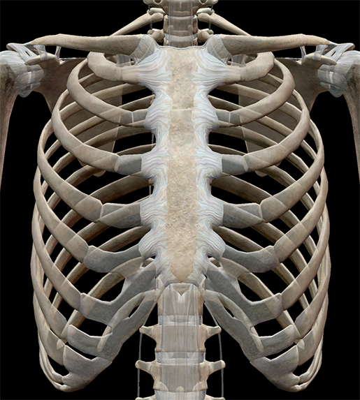 Image from Human Anatomy Atlas 2020.
2. Layers of intercostal muscles
Next, let’s check out the three layers of intercostal muscles, which are also super helpful when it comes to breathing—the external intercostals, internal intercostals, and innermost intercostals.
During normal inhalation, the external intercostal muscles contract (along with the diaphragm), elevating the ribs. You can see in the image below that their striated fibers are directed obliquely downward and laterally on the back of the thorax and downward, forward, and medialward on the front.
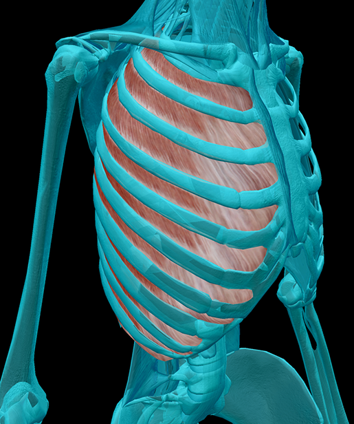 Image from Human Anatomy Atlas 2020.
The fibers of the internal intercostals run in the opposite direction to those of the external intercostals. These muscles contract to depress the ribs during forced exhalation (whereas during normal exhalation, the external intercostals just relax).
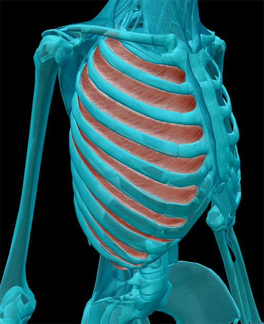 Image from Human Anatomy Atlas 2020.
The innermost intercostals help out the internal intercostals during forced expiration, and they also help stabilize the thoracic wall. Their fibers are oriented parallel to those of the internal intercostals. Additionally, nerves and blood vessels run in the space between the internal and innermost intercostals.
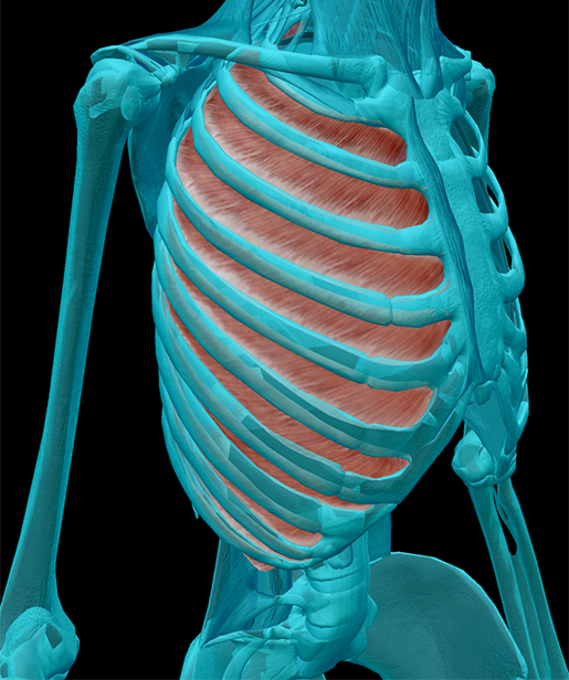 Image from Human Anatomy Atlas 2020.
3. Pleurae and pulmonary ligaments
The pleurae form a double-layered serous membrane around the lungs. The outer pleura of each lung is called the parietal pleura. It covers the mediastinum, the superior part of the diaphragm, and the inside of the thoracic wall.
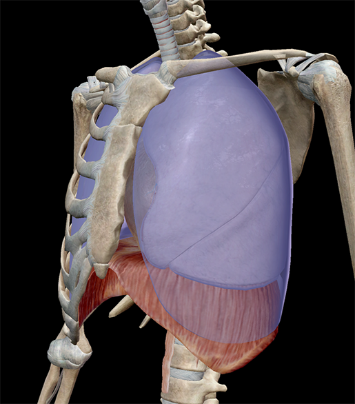 Image from Human Anatomy Atlas 2020.
The inner pleura is called the visceral pleura, and it covers the surface of each lung as well as the fissures between the lobes of each lung.
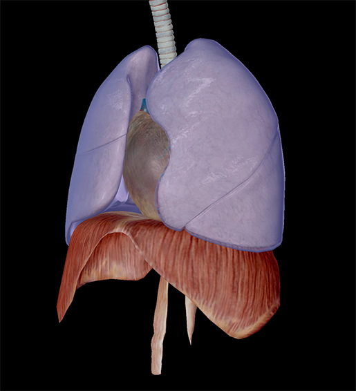 Image from Human Anatomy Atlas 2020.
The space between the parietal and visceral pleurae is called the pleural cavity, and it’s filled with a special fluid that helps prevent friction as the lungs move against the walls of the thoracic cavity.
The pulmonary ligaments are mesenteric folds that form where the parietal and visceral pleurae meet. These ligaments help keep the lower parts of the lungs in position. You can see in the image below that the shape of the left pulmonary ligament is different from the shape of the right one.
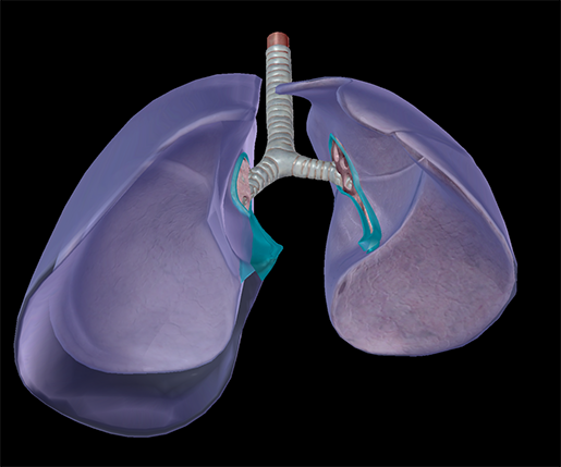 Image from Human Anatomy Atlas 2020.
4. Dissecting lungs by segment
In Human Anatomy Atlas 2020, each of the lungs is divided into 10 bronchopulmonary segments (in addition to their respective lobes).
What is a bronchopulmonary segment, you ask? Bronchopulmonary segments of the lungs are separated from one another with connective tissue, and each one has its own artery, vein, and segmental or tertiary bronchus. This means that each segment is its own anatomical unit, and one of these segments can be surgically removed without damaging or affecting the function of the others.
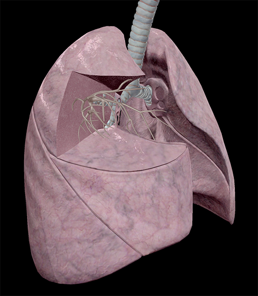 Image from Human Anatomy Atlas 2020.
It should be noted that although our model shows 10 segments in the left lung, the number of segments in an actual human body varies between 8 and 10 because some segments can be fused together.
So there you have it—an even more detailed thorax model, now available for you to explore!
You can check out these updates in video form here:
Make sure to keep an eye out for blog and video content highlighting the other exciting updates we’ve made for Human Anatomy Atlas 2020. (The best way to do that is to subscribe to the blog!)
Be sure to subscribe to the Visible Body Blog for more anatomy awesomeness!
Are you an instructor? We have award-winning 3D products and resources for your anatomy and physiology course! Learn more here.

