Five Amazing Views in Augmented Reality: Kidney and Urinary Anatomy in AR
If you want to understand what an anatomical structure does, you need to know more than just its location. You also need to comprehend its layers and facets, as well as the environment in which it exists. Textbooks, plastic models, and videos all help convey information, but if you want to really ramp it up, you can give your students a simulated cadaver lab experience!
This article shows how your students can use the new augmented reality feature in Human Anatomy Atlas 2018 on iPad/iPhone for iOS 11 to conduct their own cadaver labs by bringing 3D anatomical models into their own environment. To demonstrate this transformative learning experience, we’re going to show you how AR Mode can enhance a lesson about the kidneys and their role in the urinary system—taking it from flat 2D to interactive 3D!
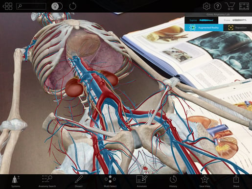
1. Put the virtual cadaver on your real lab table.
Without the urinary system we’d be (very soggy) toast, but answering questions about the urinary system’s context and how each of its individual components contribute can be difficult. Using AR, you can show your students exactly where each component of this vital system is situated, what organs surround and interact with it, and how the organ’s internal structures contribute to the blood filtration and urine creation system!
 AR from Human Anatomy Atlas.
Once AR Mode is enabled, students can lean over the virtual cadaver and study the position of the kidneys. If they want to see it from other angles, they can do so by selecting the “Prone” option to turn the cadaver over for a posterior view. Students can then hide the muscular system to see how the right kidney is slightly lower than the left.
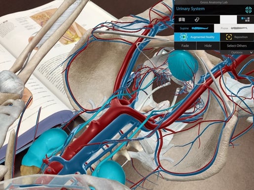
2. Check out the length of the ureters and understand their retroperitoneal location.
The ureters are about a foot long, and students can use their phones to scan the length of them while in AR Mode. Observe the location of the ureters in relation to the abdominal and pelvic spaces. It might help to have students use rulers—on themselves and on the AR model—to get a better idea of how many different systems and regions the ureters cross.
 AR from Human Anatomy Atlas.
3. Learn the structures of the kidneys.
Have your students zoom into one of the kidneys and then use the dissect feature to hide the front of the kidney; this will allow them explore the structures inside. Have them locate the renal pyramids and renal pelvis. By selecting each structure, they can read detailed definitions and hear audio pronunciations.
 AR from Human Anatomy Atlas.
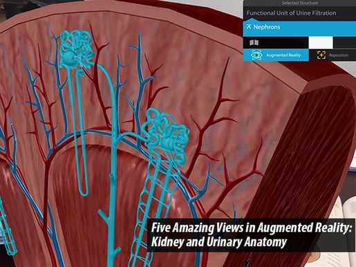
4. Understand the complexity of nephrons.
Move from a full cadaver to a specific specimen! Have students browse the microanatomy section of the app for the nephron 3D model.
Nephrons are the basic functional component of the kidneys. Now there’s a sample nephron on the virtual lab table in front of your students! Have them locate the dense glomeruli, which receive blood from the afferent arteriole and drain blood into the efferent arteriole. Next, ask them to find the peritubular capillaries and follow the path of the capillary to the vasa recta.
 AR from Human Anatomy Atlas.
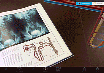
5. Follow the path of urine creation.
From the Microanatomy menu, have students select the simplified nephron model and use AR Mode to put it on their table.
Each kidney contains over a million nephrons. Students can follow the path that urine creation takes: Filtrate in the collecting duct becomes urine, which is excreted through the renal pelvis and into the ureter. Students can then return to the full cadaver model to revisit the path urine takes as it leaves the kidneys and the body.
 AR footage from Human Anatomy Atlas.
AR Mode allows students to interact with the body as if they were in a real cadaver lab—no gloves or lab coats needed!
Be sure to subscribe to the Visible Body Blog for more anatomy awesomeness!
Are you an instructor? We have award-winning 3D products and resources for your anatomy and physiology course! Learn more here.
Additional Sources:

