Posted on 12/21/16 by Sofía Pellón
We spoke to a few students who use our 3D anatomy app Human Anatomy Atlas, and they have a lot of cool insights into how using our app helps them study and learn! As you’ll learn in this blog post, Kathryn, Brandon, and Kiko each have a different perspective on learning and teaching anatomy.
Kathryn R. is a nursing student at Salem State University who also tutors undergraduate students studying Anatomy & Physiology. Brandon B. is a first-year medical student at Northeast Ohio Medical University. Kiko S. works as an adaptive yoga instructor and is currently in the process of applying to an Occupational Therapy graduate program. A former 3D Draft and Design engineer, he's currently a student at Augusta University.
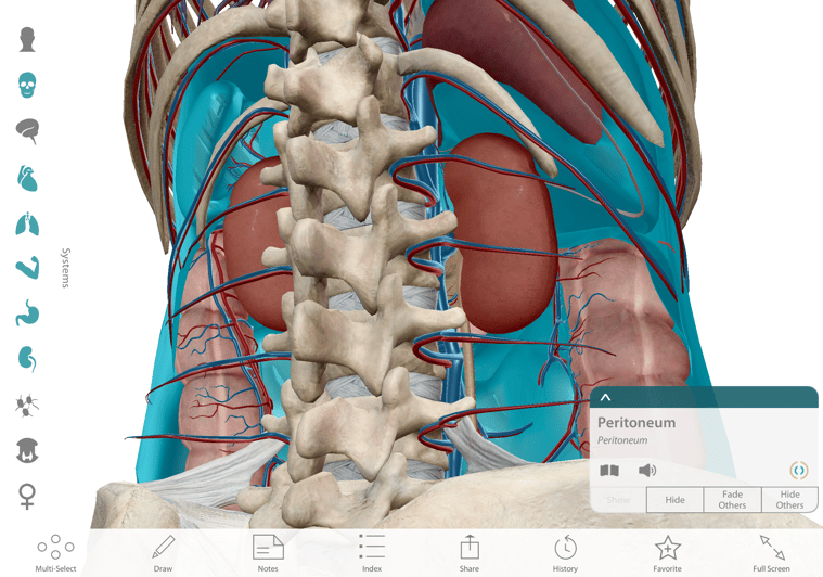
Kathryn: I think the bones and muscles in the app are very helpful to study and learn. Atlas really helps to explain what the peritoneum is because most students can't visualize what it is. It definitely helped me!
 Image from Human Anatomy Atlas.
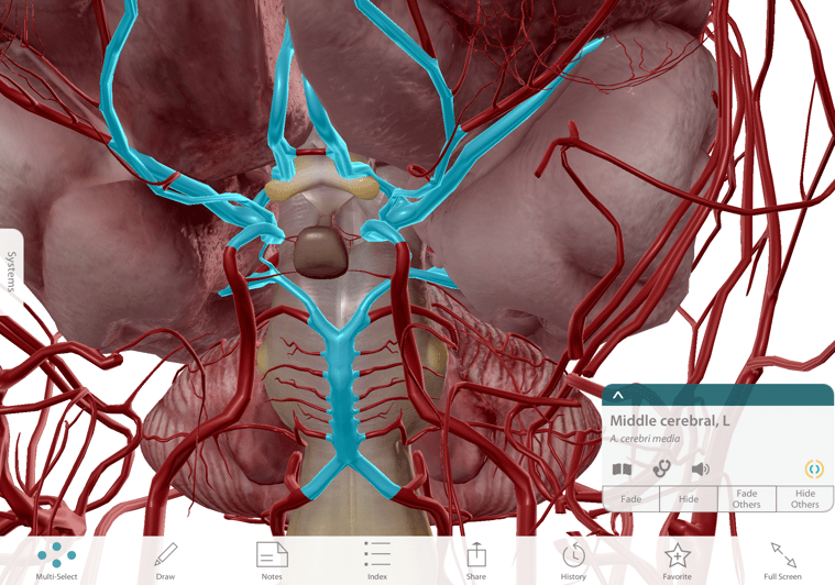
Brandon: Topics that have really resonated with me in my use of [Atlas] have included the Circle of Willis, cranial fossa and cranial nerves, brachial and lumbosacral plexuses, the shoulder and pelvic girdles (e.g., ligaments; muscle; muscle actions), and the cubital and popliteal fossae, just to name a few... [Atlas] has made understanding the muscles involved in coordinated muscle movements, as well as their respective origins/insertions, innervations, and blood supplies, much more viable and manageable.
 Image from Human Anatomy Atlas.
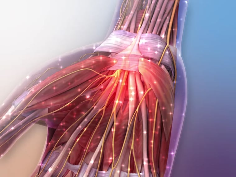
Brandon: Some of the clinical correlations have been intriguing and continue to help me put anatomical structures contributing to these conditions into perspective (e.g. Alzheimer's disease, carpal tunnel syndrome, sciatica, [etc.]). I have also enjoyed the patient education videos, such as the one on tissue repair, which efficiently clarified initial confusion in the steps of the immune response and then provided a nice summary later on in my review.
 Image from Human Anatomy Atlas.
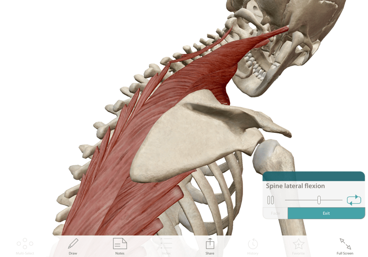
Kiko: For one, a lot of my more athletic private clients deal with bursitis, to some capacity. This is a topic that I found much more enlightening through [Atlas], as far as diagnosis and rehabilitation were concerned. Atlas made it very easy for me to show my clients what exactly their bursae were and how they were a player in their dysfunction. Another great example – anatomical movements – comes with the new Atlas 2017, which has some amazing additional features. It's so easy to get confused with flexion and extension, protraction and retraction, abduction and adduction, and all the others. With the Atlas 2017, there's a guide with interactive videos of the isolated types of movement for each joint and the muscles involved, which helped a lot with my own confusion, and also helped for cueing students and clients.
 Image from Human Anatomy Atlas.
Whether it’s visualizing the peritoneum, wrapping your mind around the Circle of Willis, or developing an intuitive understanding of abduction versus adduction, learning about anatomy presents some unique challenges! We hope these insights from Kathryn, Brandon, and Kiko have given you some great ideas for how learning in 3D can meet those challenges and get students excited about anatomy.
If Human Anatomy Atlas is helping you learn or teach something in a new way, we want to hear about it! Please feel free to let us know in the comments.
Be sure to subscribe to the Visible Body Blog for more anatomy awesomeness!
Are you an instructor? We have award-winning 3D products and resources for your anatomy and physiology course! Learn more here.
When you select "Subscribe" you will start receiving our email newsletter. Use the links at the bottom of any email to manage the type of emails you receive or to unsubscribe. See our privacy policy for additional details.
©2026 Visible Body, a division of Cengage Learning