Skin and the Physiology of Touch
Many people believe there are five senses, but it turns out that it’s a little more complicated than that. After all, our skin can also sense things like temperature, which makes the sense of touch more of a category.
Skin, the body's largest organ, helps make some of these "extra" senses possible! Today on the Visible Body, we’ll talk about skin, touch, temperature, and mechanoreceptors.
Skin Anatomy 101
Skin is made up of three main layers.
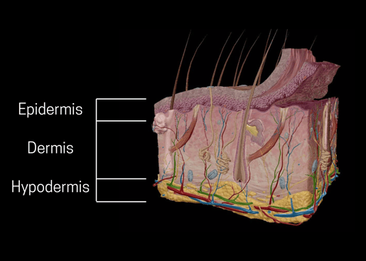
The three layers of skin, from Visible Body Suite.
The epidermis is the thin, surface layer of the skin. It's made up of several smaller layers that work together to rebuild the skin. In addition to touch receptors, the epidermis contains melanin (pigment that absorbs ultraviolet rays) and keratin, a protein that protects skin and tissue.
The layer below the epidermis is the dermis, a thick layer made up of connective tissue rich in collagen and elastin. The dermis:
- Stores water
- Regulates body temperature
- Regulates vitamin D production
- Cushions the body
- Supplies blood to the epidermis
Last but not least, the hypodermis layer is composed mostly of fatty tissue and collagen-rich connective tissue. The hypodermis:
- Separates muscle from skin
- Stores fat
- Conserves body heat
Receptors 101
This blog post focuses on the skin, but the body has many different cells that respond to stimuli. These receptors can be classified in several ways.
One way to classify receptors is to do it by type:
- A receptor cell that is specialized for a specific kind of stimulus. For example, photoreceptors contain structures that respond to light.
- A neuron with an encapsulated ending that increases the sensitivity of the nerve endings.
- A neuron with a free nerve ending, where the dendrite is unencapsulated. These are the most common nerve endings in skin.
Neuron model from Visible Body Suite.
Another way is to do it by location:
- Exteroceptors are close to the external environment, like those in skin.
- Interoceptors are close to internal organs.
- Proprioceptors are close to moving body parts, like muscles, and interpret stimuli from the movement of tissues.
Lastly, another way is by the type of stimuli they interpret:
- Chemoreceptors interpret chemicals, as I’m sure you could have guessed.
- Osmoreceptors respond to concentrations of body fluids.
- Mechanoreceptors respond to pressure, vibration, sound, and balance.
- Thermoreceptors respond to temperatures above or below normal body temperature.
The Skin’s Mechanoreceptors
There are many ways to talk about receptors, but since we’re talking about touch, let’s focus on the mechanoreceptors of the skin.
Merkel cells, Meissner's corpuscles, and Pacinian corpuscles in Visible Body Suite.
Mechanoreceptors in the skin detect different types of stimuli.
- Merkel cells, which are receptors for intensity, detect edges and points. They are located in the upper layers of the skin, and the fingertips and lips are dense with Merkel cells.
- Meissner’s corpuscles, which are receptors for velocity, detect skin motion. They are located in the dermis and project into the epidermis.
- Pacinian corpuscles, which are receptors for vibration, are found deep in the dermis.
- Ruffini endings, which detect skin stretch, temperature, and joint activity. That’s right, Ruffini endings can also act as thermoreceptors! They are located deep in the epidermis.
- There are also hair follicle receptors, which detect when the hair is moved.
Each receptor has a receptive field, an area where it responds to stimulus. Receptive fields vary by receptor, but they also vary by region of the body. Larger fields are less precise than smaller ones. Receptive fields in the fingers are smaller than receptive fields in the leg, which makes it easier to locate objects with our fingers than with our legs.
Slow adapting receptors continue to fire while a stimulus is present, while fast adapting fibers only fire when something changes. As such, slow adapting receptors like Merkel’s disks are good at detecting shape and pressure while fast adapting receptors like Pacinian corpuscles are good at detecting movement and vibration.
The Path of Sensory Information
Ion channels are proteins located in the plasma membrane of a cell. They allow specific ions to pass in or out of the cell.
In response to stimulus, ion channels in cells open up, generating electrical signals. Different ion channels respond to different types of stimuli, like different temperatures and types of pressure or movement. For example, the ion channel PIEZO1 responds to touch, and PIEZO2 senses touch and also conveys information about the body, like how we can tell when our muscles are stretching and when the lungs are fully inflated.
When the ion channels respond to stimulus, the receptors send signals to neurons, which send the message to the brain to be processed. The spinal cord processes sensory information if quick action is needed—for example, if you put your hand on a hot iron and need to quickly pull it away.
When the message reaches the brain, the sensory cortex processes the information. To be specific, the information is processed in the somatosensory cortex, which is made up of Brodmann’s areas 1, 2, and 3. 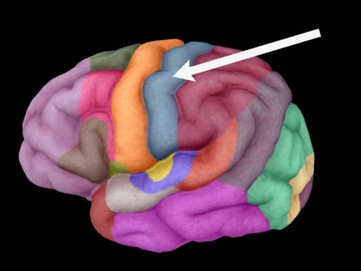
Arrow pointing to primary somatosensory cortex. Image from Visible Body Suite.
Conclusion
Different kinds of stimuli trigger different kinds of receptor cells in the skin by activating ion channels on the cell's surface. Different ion channels and different kinds of receptor cells (like Merkel cells, Meissner's corpuscles, Pacinian corpuscles, and Ruffini endings) respond to different kinds of stimuli, like temperature change, vibration, pressure, and motion. The receptors send an electrical impulse to the brain, where the sensory information is processed.
Want to learn more? Check out these other blog posts on the integumentary system, the senses, and more!
- Getting An Earful: The Anatomy Behind Hearing and Balance
- Let's Untangle This: All About Hair
- Marvelous Melanin: A Pigmentation Primer
- All About Itching
Be sure to subscribe to the Visible Body Blog for more anatomy awesomeness!
Are you a professor (or know someone who is)? We have awesome visuals and resources for your anatomy and physiology course! Learn more here.

