Osteon Powers: A Dive into Compact Bone’s Functional and Structural Unit
Today on the Visible Body Blog, we’re talking about osteons! As the functional and structural unit of compact bone, the osteon is responsible for the strength of the bone, the influx of nutrients into the bone, and waste removal from the bone.
Compact vs. spongy bone
Before we discuss the details of the osteon, let’s first talk about compact bone!
There are two types of bone tissue: spongy and compact.
Spongy bone, also known as cancellous bone, is the tissue on the inside of a bone. It is made up of slender fibers and lamellae and is filled with marrow, which creates red blood cells. Spongy bone adds flexibility and a level of shock absorption to the bone, and it also stores minerals. Bones store 99% of the body's calcium!
On the other hand, compact bone is the dense bone tissue on the outside of the bone. It’s covered in the periosteum, a membrane that covers the entirety of the bone and is where muscles and tendons attach.

Compact bone highlighted on Human Anatomy Atlas 2022 +'s sectioned femur model.
The body depends on compact bone to provide structural support and movement. Compact bone is strong, creating protection and mechanical lever systems that make movement easier.
What is an osteon?
Osteons are the functional units of compact bone. The term functional unit means the smallest unit that can carry out one of the organ’s functions. Compact bone is made of cylindrically-shaped osteons connected together.
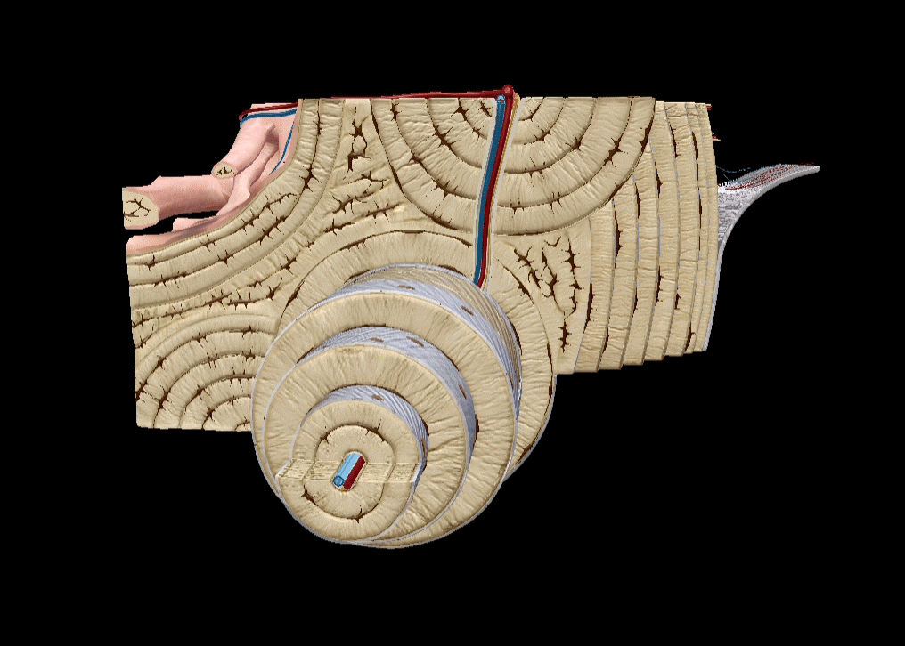
Osteon GIF from Human Anatomy Atlas 2022 +.
Though people like ”Father of Microbiology” Antonie van Leeuwenhoek observed some parts of osteons, Clopton Havers was the first to study osteons in depth, publishing the first description of an osteon in his 1691 book, Osteologia nova. This is why osteons are sometimes called Haversian systems.
Put simply, the osteon is made of lamellae, lacunae, and osteocytes arranged around a central canal. Let’s dive deeper!
Lamellae
It’s the extracellular matrix around the cells that gives compact bone its hardness and rigidity. This matrix is made of both organic and inorganic materials. For example, collagen provides tensile strength and hydroxyapatite crystals provide the bone with compressive strength.
Lamellae (singular: lamella) are layers of matrix.
There are three types of lamellae.
- Interstitial lamellae are irregularly shaped and fill in the spaces between osteons
- As their name suggests, circumferential lamellae wrap around the circumference of the entire bone
- Concentric lamellae are circular, arranged concentrically around the central canal of each osteon
Interstitial, circumferential, and concentric lamellae. GIF from Human Anatomy Atlas 2022 +.
Lacunae and osteocytes
Lacunae (singular: lacuna) are gaps or empty spaces. Lacunae within the lamellae contain osteocytes.
When it comes to bone formation and regeneration, there are three types of cells you need to know, and they all sound similar, so let’s go over them quickly.
Osteoblasts build bone by secreting extracellular matrix. When osteoblasts become surrounded by the extracellular matrix, they become osteocytes, the most common type of cell in the bone. Osteocytes have access to blood vessels and provide the bone with nutrients. Osteoclasts, on the other hand, break down matrix and secrete the resulting products into blood.
Located in lacunae, osteocytes perform a multitude of functions, such as
- Controlling osteoblast and osteoclast activity
- Helping metabolize phosphates
- Exchanging nutrients and wastes with other cells
- Preserving the matrix
- Regulating bone mass
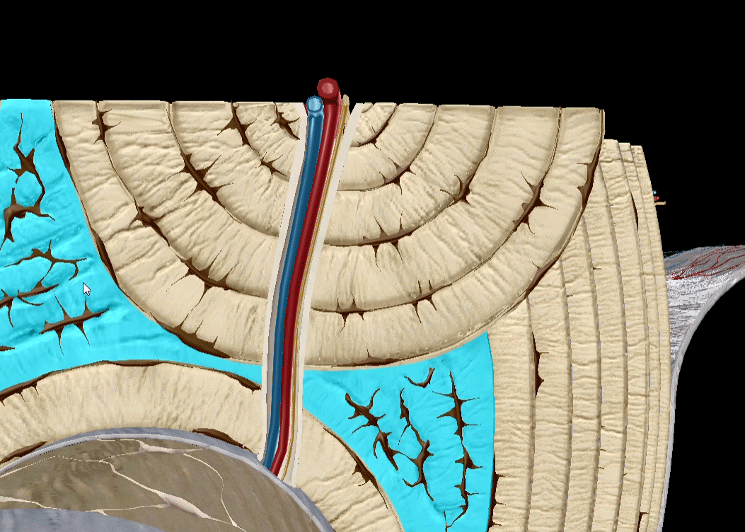
Canals
Osteocytes are located within lacunae, which are in turn located in lamellae. The concentric lamellae are arranged around the central canal, also known as the Haversian canal.
Each central canal runs parallel to the long axis of the bone. It’s where blood vessels, lymph vessels, and nerves are located. It also contains a small amount of connective tissue.
Perforating canals, also known as Volkmann’s canals, run at right angles to central canals. Perforating canals connect the central canal’s blood vessels with the periosteum’s blood supply.
.jpg?width=515&name=My%20project%20(6).jpg) Central and perforating canals in Human Anatomy Atlas 2022 +.
Central and perforating canals in Human Anatomy Atlas 2022 +.
Conclusion
Want a quick review of the structure of bone tissue? Check out this video!
To wrap it all up, osteons are structural and functional units of compact bone. Every osteon is made up of a central canal (which contains nerves and blood vessels), perforating canals, lamellae (configurations of bone matrix), and osteocyte-containing lacunae (holes). All of these are arranged concentrically around the central canal.
Be sure to subscribe to the Visible Body Blog for more anatomy awesomeness!
Are you an instructor? We have award-winning 3D products and resources for your anatomy and physiology course! Learn more here.

