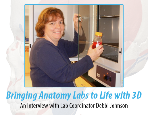
Bringing Anatomy Labs to Life with 3D: An Interview with Lab Coordinator Debbi Johnson
In the A&P lab at Western Connecticut State University, there is all the great stuff you’d expect:
Microscopes. Check.
Plastic legs and arms hanging in a glass-fronted cabinet. Check.
A painted cross section of a kidney mounted on a piece of wood.
A heart in a jar.
Some skeletons ready for felted muscles to be attached to bones.
Check, check, check.
But the students also have a 3D human body available, right at their fingertips on computers, phones, and tablets. Spring semester was the first time Lab Coordinator Debbi Johnson’s students had Visible Body Suite (previously called Human Anatomy Atlas) at their disposal. And it made a huge difference, especially for learning origins and insertions.
“My students loved it for bones and muscles,” Johnson says. “I saw a huge improvement in their knowledge of these structures. They did better in labs and on quizzes after using [VB Suite]. They kept saying, ‘Now I get it!’”
What’s the magic behind this uptick in performance? Using VB Suite, they practiced at home after lab and could better prepare for the quiz. “They know,” she says, “whatever they did this week is on next week’s quiz.”
Take muscles, for example. For the quiz, Johnson provides a chart for origins and insertions. In their own words, students must list the origin, insertion, and the action for each muscle. They use the plastic models that have each structure numbered to guide them through the exercise.
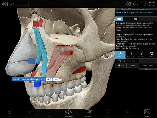
Pictured: The attachment points of the levator labii superioris alaeque nasi in VB Suite.
“Using [VB Suite] before the quiz, they needed to show us they knew where each muscle was, not just its name,” she says. The ability to see the text of origins, insertion, action, innervation, and blood supply, in addition to the actual pins and paint, nerves, and vasculature, made an impact. As did VB Suite's bank of animated muscle actions.
“That they could watch the muscle move focusing on just the leg, just the arm—this made a huge difference. I never heard a student say they got that concept before. Never.”
Joanna P., a sophomore in Johnson’s class and an aspiring speech pathologist, used VB Suite recently to get ready for the origins and insertions activity. “For the muscles, that was the really hard one. I had to make sure I could label muscles properly using Visible Body. I took a copy of the chart we use for origins and insertions and made sure I could fill it all in.”
Joanna says the immediacy of learning the answer to a structure name made a big difference in preparing for quizzes. “I like to quiz myself to see if I know it. With diagrams, I can’t see all of the structures as easily. With Visible Body, I just click or tap to see what it is.”
The ability to rotate the body in 3D and to zoom in and out brings concepts to life, instead of viewing them flat in a book. “You can move everything, which is amazing,” Joanna says. “Three dimensional instead of just seeing lines on a diagram in a book is great.”
Freshman Kassidy C., who was just accepted into the nursing program at Western Connecticut State, uses VB Suite on her iPad—in particular to prep for exams. “Especially in my first semester, I had a little trouble learning the bones. I could memorize the names and general locations of the bones and their connections to each other. However, Visible Body allowed me to properly visualize and learn their exact locations in the body and relationship with other bones,” she says. “This was, obviously, very important for my lab practical exams.”
Pictured: Interacting with the disarticulated skull 3D model in VB Suite.
For Kassidy, VB Suite is clean and well organized, with plenty of helpful details. “My favorite feature is the ability to isolate systems or layer others on top of each other,” she says. “It’s one thing to get to know a system but another thing to see the relationship between multiple ones.”
Johnson projects VB Suite from the front of the room to the class while discussing the circulatory system before the identification activities begin. “I show them you can hide or fade a bone, and isolate just the arm while you’re learning the arm vasculature.”
For the circulatory system unit that they just finished, the students got to tap on each and every blood vessel on the provided list without leafing through pages in their lab manual to correlate the list.
“We give [the students] a list of what to ID, all the arteries and veins; knowing them preps [the students] for a lab activity,” Johnson says. “I showed them the app, and that all of the blood vessels were there. Those [students] who used it said it really helped them.”
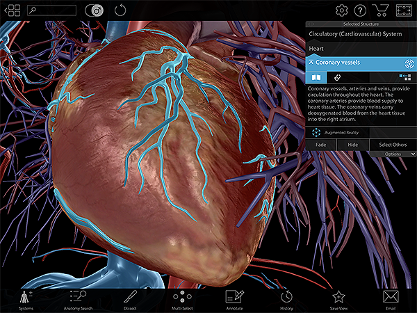
Pictured: The coronary vessels (highlighted in blue), from VB Suite.
The lab activity involves a numbered plastic model—students have to write the name of the vessel next to the number on their worksheet. Prepping for the hundreds of structures can be a bear, Johnson says. “But knowing the answer immediately using [VB Suite] helps. If you just have a picture in a book, you cover up the answers with your hands. And you can sort of cheat because you can look ahead to what the next one is. You’re not really learning it that way. With [VB Suite], the answer is there, then it’s not there. It’s instant, but the student isn’t getting the next answer.”
Johnson can assign sections of the circulatory system bit by bit, so students can master one region at a time and build on that knowledge. “There’s the chest, the neck, and a few head structures they need to learn that night, then tomorrow we’ll look at the arm, then look at abdomen, then the leg,” she says. “Then they can start putting two together, so by the next week they’ll have them all memorized.”
Want more?
- Learn more about how and why students use 3D anatomy apps in their studies.
- Read our interview with Ph. D student Tim Fleischer, who explains how 3D apps improve student retention.
- Check out Visible Body's anatomy apps and find out how to bring 3D anatomy to your classroom or lab!
- Never miss a thing! Sign up to get our awesome emails and be the first to know about free anatomy content, new blog posts and eBooks, secret sales, and more!

