Anatomical Variations of the Circulatory System
The human body doesn’t always look like it does in textbook illustrations. The term anatomical variant refers to deviations from the norm that don’t impair functions, though some anatomical variants can cause symptoms. These are also known as normal variations.
In recent decades, the development and rise of imaging techniques has uncovered more and more variants—for most people, anatomical variations go undetected until they are uncovered by diagnostic imaging or during a post-mortem examination.
The circulatory system has the most known variations, and arterial variations are the best-described of the anatomical variants. After all, the average adult’s circulatory system is more than 60,000 miles long—there’s a lot of room for variation!
Today on the Visible Body blog, we’ll discuss some variations of the circulatory system.
Aortic Arch
Before we talk about variants of the aortic arch, let’s do a quick review of the aorta itself.
At just over an inch in diameter, the aorta is the largest artery in the body. It has three main sections: the ascending aorta, the aortic arch, and the descending aorta.
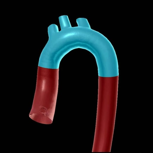
Aortic arch highlighted. Photo from Human Anatomy Atlas 2022 +.
The ascending aorta is about two inches long and rises up from the heart’s left ventricle. The left and right coronary arteries branch from the ascending aorta to supply the heart with blood.
The aorta then curves over the heart in a candy cane shape: this is the aortic arch. Three branches come off of your average aortic arch: the brachiocephalic (which branches into the right subclavian and the right common carotid arteries), the left common carotid, and the left subclavian arteries.
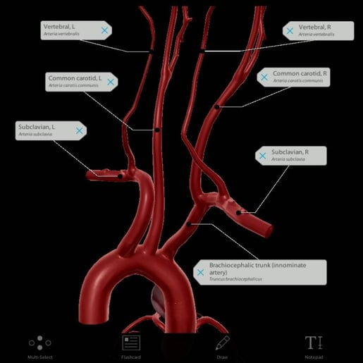
Photo from Human Anatomy Atlas 2022 +.
The descending aorta moves down the chest—in a candy cane comparison, this is the handle. In the abdomen, it branches into several arteries that deliver blood to most of the major organs.
Now let’s check out some variants of the aortic arch!
Isolated vertebral artery
In most people, the left and right vertebral arteries rise from their corresponding subclavian arteries, but in about 5% of the population, the left vertebral artery arises from the aortic arch.
The isolated left vertebral artery often enters the cervical vertebrae’s transverse foramina higher than normal, at C5 rather than C6. In about 4% of cases, the isolated left vertebral artery terminates in the posterior inferior cerebellar artery instead of merging with the basilar artery.
Bovine arch
The bovine arch is the most common aortic arch variation, occurring in about 27% of the population. It’s more common among people of African or South American descent.
Bovine arch can be divided into two subtypes.
The most common form of bovine arch (in 20% of the population) configuration is where the left common carotid artery and brachiocephalic artery share a common trunk. This is also called a bicarotid trunk or truncus bicaroticus.
.jpg?width=515&name=My%20project%20(3).jpg)
Bovine arch illustration made using the draw feature in Human Anatomy Atlas 2022 +.
7% of the population has a configuration in which left common carotid artery branches directly from the brachiocephalic trunk.
Bovine arches are usually asymptomatic, but it’s important for physicians to be familiar with these configurations, particularly when performing surgery like carotid stenting where the shape of the arch can add an extra challenge.
Fun fact: the name bovine arch is a bit of a misnomer. Cows’ aortic arches aren’t shaped that way; their brachiocephalic trunk branches in three ways into the bilateral subclavian arteries and a bicarotid trunk.
Aberrant right subclavian artery
The aberrant right subclavian artery, aka arteria lusoria, occurs in about 0.5%-2% of the population.
In this configuration, there is no brachiocephalic trunk; instead, the right and left common carotid arteries and the right and left subclavian arteries rise from the aortic arch. The right subclavian artery is the most distal and usually extends behind the esophagus to reach the right arm.
If the right subclavian artery presses against the esophagus, this can cause dysphagia lusoria, a swallowing impairment. Dysphagia lusoria is difficult to diagnose because symptoms are nonspecific, but most cases are mild.
Circle of Willis
It’s easy to think of anatomical variants as unusual, but the circle of Willis underscores that anatomical variants are more commonplace than you might think.
The circle of Willis has an extremely important role: it supplies blood to the brain. It connects the internal carotid, vertebral, and basilar arteries to the brain, and it also provides collateral circulation between the forebrain and the hindbrain.
The circle of Willis surrounds the pituitary gland and the optic chiasm. The textbook description would say that it is made up of the anterior, middle, and posterior cerebral arteries and two communicating branches called the posterior and anterior communicating arteries. Numerous branches then supply blood to different parts of the brain.
However, only 20-25% of the population has a complete circle of Willis.
The most common variation is the absence of a posterior communicating artery, but there are many different variants. Vessels can be:
- Duplicated, where two vessels fuse.
- Fenestrated, where a vessel divides and then those divisions fuse back together.
- Hypoplastic, where the vessel is underdeveloped.
- Absent, where the vessel is, well, absent.
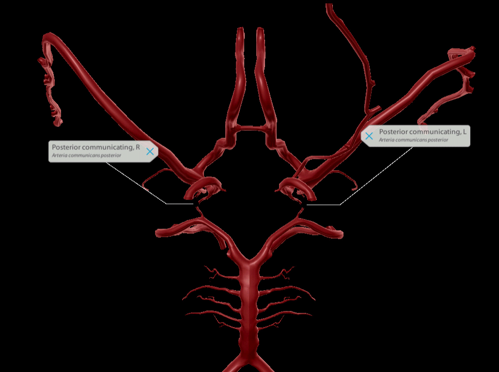
Missing posterior communicating arteries is the most common variation of the circle of Willis. GIF from Human Anatomy Atlas 2022 +.
Celiac Trunk
The typical celiac trunk arises from the aorta below the aortic hiatus at the diaphragm. It branches off into three arteries: the left gastric, the common hepatic, and the splenic. Those arteries branch off further to supply the stomach, pancreas, liver, spleen, and surrounding structures with blood. 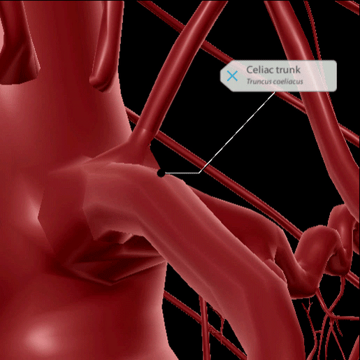
The typical celiac trunk. GIF from Human Anatomy Atlas 2022 +.
Typical trifurcation, when the arteries have a common origin, is called a true tripod. A variation called a “false tripod” occurs when one artery arises before the other two.
In about 11% of the population, the celiac trunk bifurcates instead of trifurcates. In bifurcation, the trunk splits into two arteries, one of which bifurcates again to form the third artery.
There are many different variations in branching possible—between the three arteries and all the possibilities of branching, it’s a bit of a mix-and-match situation.
Renal Artery
The renal arteries are two lateral branches from the abdominal aorta, one for each kidney. Before entering the kidney, each of the renal arteries divides into five branches that supply different segments of the kidney. The segmental renal arteries enter the kidney through the hilum, which is also where veins, nerves, and ureters enter and exit.
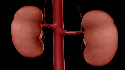
Typical renal arteries. Photo from Human Anatomy Atlas 2022 +.
Accessory renal artery
Accessory renal arteries, also known as extra renal arteries, typically arise from the aorta above or below the main renal artery. Accessory renal arteries are common, presenting in 25-30% of the population.
There are many different variations of accessory renal arteries, and they occur bilaterally in 10-15% of cases. People can have double, triple, and even quadruple renal arteries on one or both sides.
Some examples of accessory renal arteries include:
- An artery that supplies the inferior pole of the kidney
- An artery that enters through a surface of the kidney
- An artery that arises from the celiac trunk
- An artery that arises from the superior or inferior mesenteric arteries
- An artery that arises from the middle sacral artery
Accessory renal arteries do not seem to impact health, and they do not meaningfully influence clinical outcome in the case of kidney donation, though some believe that accessory renal arteries could be a cause of renin-dependent hypertension.
Early division of renal artery
The other variation in renal artery anatomy is an early division of the renal artery. In this variation, the renal artery branches into segmental arteries sooner than usual. It’s as simple as that!
Conclusion
Let’s review!
The brachiocephalic, the left common carotid, and the left subclavian arteries all branch from the aortic arch. However, the aortic arch has several variants including the isolated vertebral artery, bovine arch, and the aberrant right subclavian artery.
The circle of Willis is a circle of connected arteries that supply blood to the brain. A complete circle of Willis is only found in about a fourth of the population—there are quite a number of variations that can occur.
The typical celiac trunk branches into the left gastric, common hepatic, and splenic arteries. There are many variations in this arrangement, including a false tripod (when one artery splits before the other two) and a bifurcating trunk.
The renal arteries supply the kidneys with blood, and there is typically one renal artery per kidney. The two variations of the renal artery are accessory renal arteries, where more than two renal arteries exist, and the early division, where the renal artery branches into accessory arteries earlier.
Be sure to subscribe to the Visible Body Blog for more anatomy awesomeness!
Are you an instructor? We have award-winning 3D products and resources for your anatomy and physiology course! Learn more here.

