


3D Skeletal System: 5 Cool Facts about the Hip Bones
I know, I know, it's another blog post about the pelvic girdle. Well, sort of. Sometimes it's a thankless job, holding the weight of our upper half, so this post is going to be dedicated to my favorite pair of jutting wings—the hip bones!
So sit down and take a load off, and let's dive in!
1. The proper term for the hip bones is "os coxae."
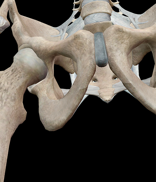
Image from Human Anatomy Atlas.
Os coxae comes from the Latin words "os" meaning bone and "coxae" from the old Latin word for hip.
2. Each hip bone is actually made up of three bones.
It may look like one bone, but each hip bone is made up of the ilium, pubis, and ischium, which are completely fused.
3. There's a cavity in each hip bone.
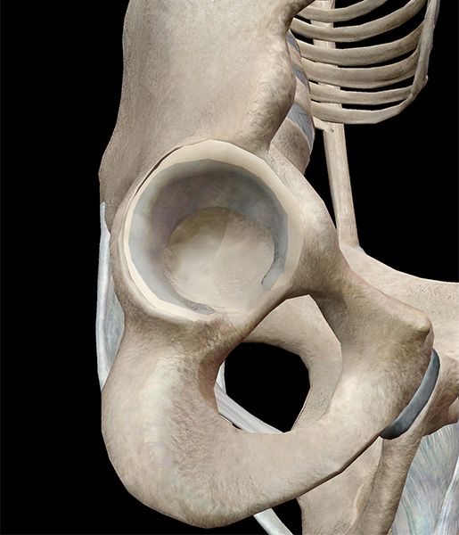
Image from Human Anatomy Atlas.
No, seriously! The concave cavity in the hip socket is known as the acetabulum, which is where the head of the femur articulates.
4. There are noticeable differences between the male and female hip bones.
The female hip bones are more delicate and shallow than the male's, with less sloped ilia. However, the superior aperture of the female pelvis is larger and more circular than the male's.
5. There is a giant hole between the hip bones.
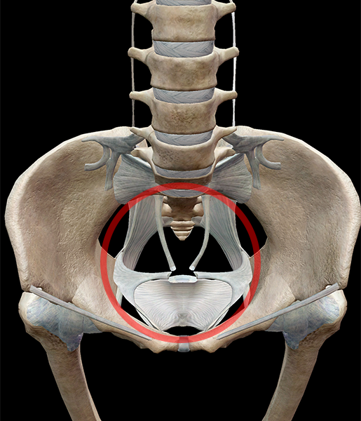
Image from Human Anatomy Atlas.
Well, technically it's a space more than a hole. The superior aperture is the space that divides the abdominal cavity from the pelvic cavity. If you look at the pelvic area from a superior view, you will see the superior aperture is circular and formed by the hip bones, sacrum, and pubic symphysis.
| Did you know? The hip bones are part of the appendicular skeleton, which also includes the shoulder girdle and the upper and lower limbs. |
Be sure to subscribe to the Visible Body Blog for more anatomy awesomeness!
Are you an instructor? We have award-winning 3D products and resources for your anatomy and physiology course! Learn more here.
Related Posts:
- 3D Skeletal System: Atlas, Axis, and the Atlanto-Axial Relationship
