

Studying the Peritoneum with VB Suite
Here at Visible Body, we are always listening to our customers’ suggestions about how to improve our apps. When students and instructors at Harvard Medical School asked if we could enhance the peritoneum structure in Visible Body Suite, our biomedical visualization team worked with them to create a more detailed peritoneum for both the male and female model.
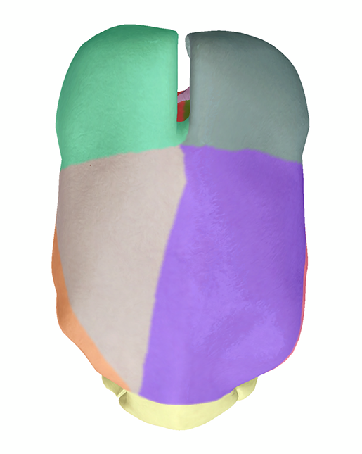
The new peritoneum landmarks view (female, anterior). Image from VB Suite.
In this post, we’re going to show you the peritoneum model in VB Suite and discuss how it incorporates structures students need to know for conducting FAST (focused assessment with sonography for trauma) ultrasound exams on patients.
What is the peritoneum?
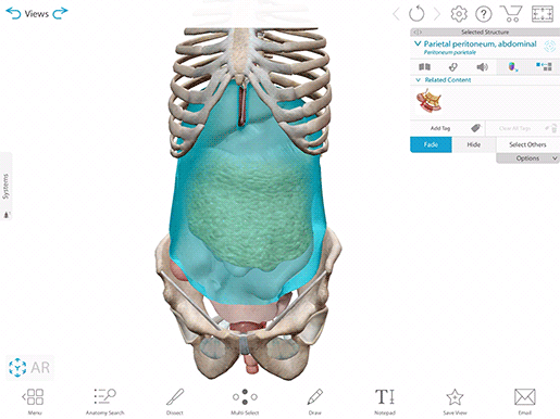
The peritoneum is the serous membrane that lines the abdominal and pelvic cavities. It has two layers: the parietal perineum, which is attached to the walls of the abdomen and pelvis, and the visceral peritoneum, which covers the internal organs. The peritoneal cavity is the fluid-filled space between the parietal and visceral peritoneum. This fluid allows the organs of the abdominal and pelvic cavities to move past one another smoothly.
 The peritoneum (male). Have VB Suite on your mobile device? See this view in 3D!
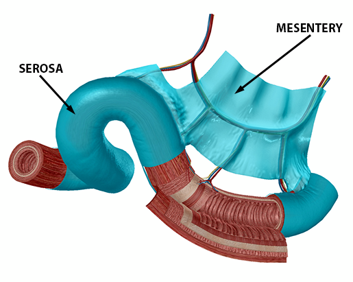
Some folds of the peritoneum are referred to as individual structures. For example, a double-fold of peritoneum forms the mesentery, which attaches the intestines to the abdominal wall and is continuous with the serosa.
Have VB Suite on your mobile device? See this view in 3D!
The lesser omentum connects the liver, stomach, and part of the duodenum. It helps hold the stomach in place and also serves as a support for blood and lymph vessels that supply the organs of the upper abdomen.
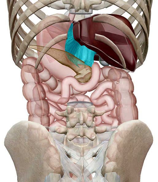
Posterior view of the lesser omentum. Have VB Suite on your mobile device? See this view in 3D!
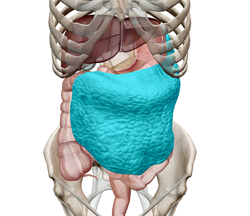
The greater omentum connects the stomach, diaphragm, transverse colon, and posterior abdominal wall. It stores adipose tissue (fat) and cushions the anterior and lateral abdominal organs.
Have VB Suite on your mobile device? See this view in 3D!
With VB Suite, you can take a more in-depth look at the regions of the peritoneum and peritoneal cavity, which you can access by highlighting the peritoneum and selecting the “landmarks” view from the info box.
 The peritoneum landmarks button. Image from VB Suite.
In order to conduct diagnostic procedures like the FAST exam, medical professionals need to be familiar with these regions—so before we go into more detail on them, let’s quickly go over what the FAST exam is and why it’s clinically important.
What is a FAST exam?
Ultrasound, also called sonography, is an imaging technique in which a technician uses a small handheld device called a transducer to aim high-frequency sound waves at an area of the body. Some of these sound waves bounce back when they hit structures inside the body, and a computer analyzes these and uses them to generate an image. One of the benefits of ultrasound is that it doesn’t involve radiation like an X-Ray or CT scan. This is why it’s the imaging method of choice for looking at developing fetuses.
The FAST exam is a specific ultrasound examination used by medical professionals to look for hemorrhage or abnormal fluid in the pericardium, pleural cavity, and peritoneal cavity after traumatic injury. The eFAST (extended FAST) exam is basically the regular FAST with additional views to examine the lungs. It’s a bedside ultrasound, which means the imaging equipment can be brought to where the patient is.
Peritoneum landmarks in VB Suite
When a FAST/eFAST exam is being conducted, there are typically a few key views that focus on a number of important structures, such as particular organs and regions of the peritoneum.
The Right Upper Quadrant (RUQ) view “uses the liver as an ultrasound window” to look for fluid in the hepatorenal space (Morison’s pouch). The right subphrenic space, which lies between the liver and diaphragm, is another place fluid can accumulate. In addition, moving the ultrasound probe towards the head (cephalad) provides a view of the right pleural space and caudal movement provides a view of the right paracolic gutter.
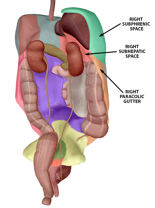 Right Upper Quadrant peritoneum landmarks (female model). Image from VB Suite.
The Left Upper Quadrant (LUQ) view uses the spleen to look at the left subphrenic space (the space between the spleen and the diaphragm) and the splenorenal recess (the space between the spleen and left kidney). The left pleural space and left paracolic gutter can also be examined via movement of the probe.
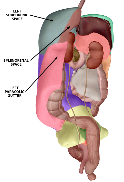
Left Upper Quadrant peritoneum landmarks (female model). Image from VB Suite.
The Pelvic view is used to examine the rectovesical recess or rectouterine recess. The rectovesical recess is the space between the rectum and the bladder in the male pelvis, while the rectouterine recess is the space between the rectum and the posterior uterine wall in the female pelvis.
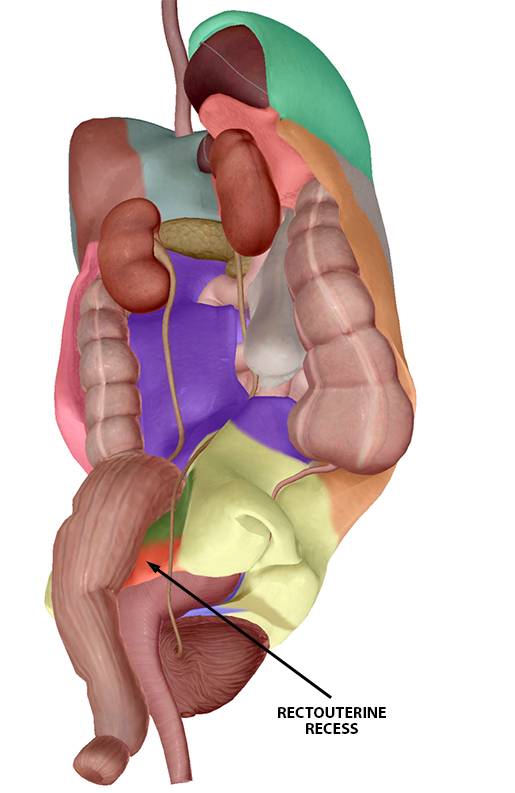
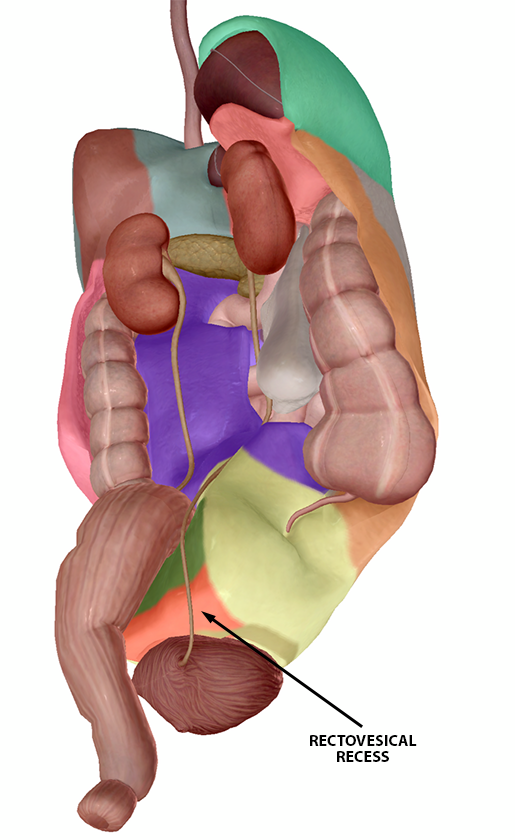
Rectouterine recess (f) and rectovesical recess (m). Images from Vb Suite.
Other FAST-related structures in VB Suite
A Pericardial (Subxiphoid, Subcostal) view is used to assess fluid buildup in the pericardium. It uses the liver’s left lobe as its ultrasound window. If the ultrasound probe is angled in a particular way, the inferior vena cava and hepatic veins can also be examined.
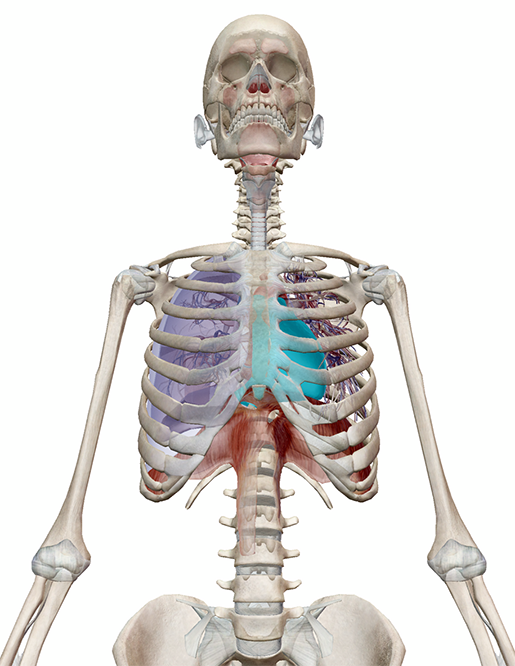
The pericardium (highlighted). Have VB Suite on your mobile device? See this view in 3D!
An Anterior Thoracic view (part of eFAST or extended FAST) can be used to look at the pleural spaces. The pleural spaces can be examined from various points on each side of the chest, and such examination is important for diagnosing pneumothorax. (See this post on the Deadly Dozen for the specs on that!)
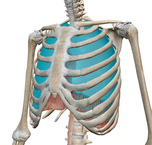
The pleurae (highlighted). Have VB Suite on your mobile device? See this view in 3D!
We hope you’ve enjoyed this lightning-fast overview of the structures evaluated using the FAST exam! The peritoneum may not get a lot of glory, but it’s important to know its different regions when it comes to diagnosing and treating injuries.
Be sure to subscribe to the Visible Body Blog for more anatomy awesomeness!
Are you an instructor? We have award-winning 3D products and resources for your anatomy and physiology course! Learn more here.
Additional Sources:
