


Learn Muscle Anatomy: Knee Joint Group
As I sit here, typing, I've got my legs crossed. My chiropractor would probably throw something at my head if he knew. Oh well. Is YOLO still a thing? Because YOLO.
Where my legs are crossed (at the knees) there are several muscles of the lower limbs in play. And of course there are—do you know how many anatomical structures it takes to move your knee? Probably more than you think.
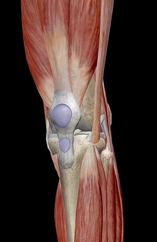
Image from Muscle Premium.
Muscles of the Knee Joint
The muscles of the knee joint are incredibly important. They move when you do—when you walk, run, dance, stretch your legs, or make any action you can think of that involves bending the knees.
There are two muscle groups that act on the knee joint: the quadriceps femoris and the posterior compartment of the proximal leg. In addition to these groups are the plantaris, articulus genu, semiteninosus, semimembranosus, and popliteus.
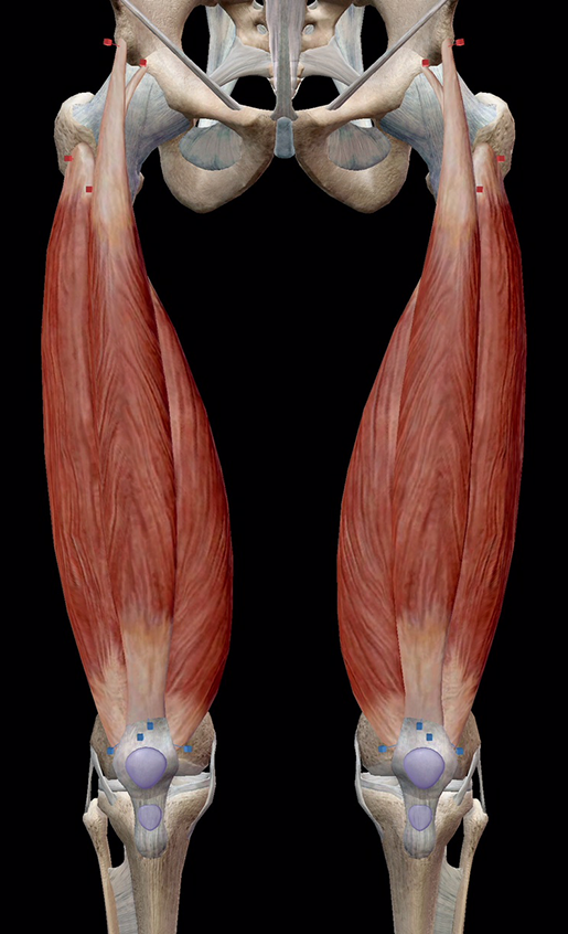
Image from Muscle Premium.
Let's take a look at the quadriceps femoris group.
Quadriceps Femoris Group
|
Muscle |
Attachments |
Actions |
|
Rectus femoris |
Originates on the anterior inferior iliac spine and a groove superior to the acetabulum; inserts on the common tendon of the quadriceps enclosing the patella, and on the tibial tuberosity |
Extension of the leg at the knee joint; flexion of the hip |
|
Vastus lateralis |
Originates on the greater trochanter and upper lateral surface of the linea aspera; inserts on the patella via the quadriceps tendon, and the tibial tuberosity via the patellar ligament |
Extension of the leg at the knee joint |
|
Vastus intermedius |
Originates on the upper two-thirds of the anterior and lateral surfaces of the femur; inserts on the common tendon of the quadriceps enclosing the patella, and on the tibial tuberosity |
Extension of the leg at the knee joint |
|
Vastus medialis |
Originates on the intertrochanteric line and medial lip of the linea aspera; inserts on the common tendon of the quadriceps enclosing the patella, and on the tibial tuberosity |
Extension of the leg at the knee joint |
Bursae
The body employs a bunch of different anatomical structures to keep friction down, and bursae are one of them.
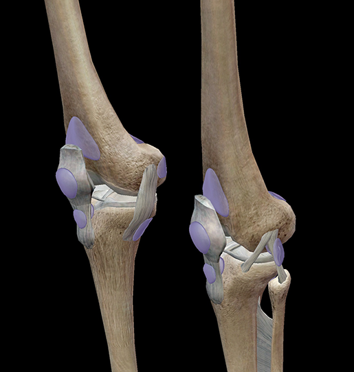
Image from Muscle Premium.
Bursae are fluid-filled sacs that can be found anywhere skin rubs over bone, and where a muscle, ligament, or tendon glides directly over the periosteum (outer surface) of a bone. The synovial fluid in the bursae linings provides lubrication, enabling freedom of movement between contiguous connective tissue surfaces.
The bursae found in the knee include the superficial prepatellar, superficial and deep infrapatellar, medial and lateral gastrocnemius, suprapatellar, and quite a few more.
Knee Extension
So, I'm still sitting with one knee crossed over the other and somewhere my chiropractor just became flushed with rage and has no idea why. If I stand, however, I will be straightening, or extending, my knees.
Extension increases the angle between body parts. Flexion decreases the angle. Stand up for a moment and keep your legs perfectly straight—this is extension.
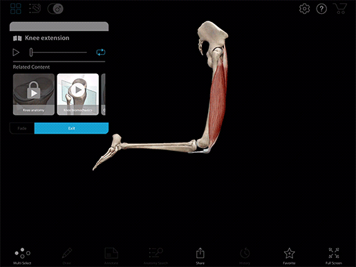
Video footage from Muscle Premium.
The angle is around 180 degrees. Now, stand on one leg and lift the other until it's bent at the knee—this is flexion.
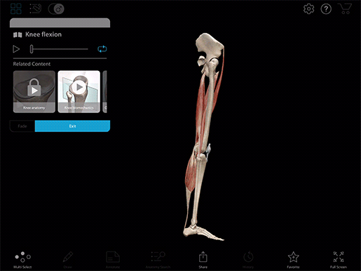
Video footage from Muscle Premium.
And now I'm going to give my chiropractor's blood pressure a rest and put both my feet on the floor.
Want to learn more about how knee flexion works? Check out our Knee Flexion eBook!

Be sure to subscribe to the Visible Body Blog for more anatomy awesomeness!
Are you a professor (or know someone who is)? We have awesome visuals and resources for your anatomy and physiology course! Learn more here.
Related Posts:
- Learn Muscle Anatomy: Serratus Posterior Superior and Inferior
