

DNA and RNA Basics: Replication, Transcription, and Translation
DNA (deoxyribonucleic acid) is one of the most important molecules in your body, and though around 99.9% of your DNA is the same as that of every other human, the 0.1% that’s different is what makes you genetically unique! This tiny biological structure is the ultimate instruction manual, containing the “recipes” for the proteins your body needs to develop and function.
Today, we’re going to give you a primer on the basics of DNA. We’ll talk about its structure, how it replicates, and the role it plays in the production of proteins.
March 2022 Update:
Visible Biology is now available! Visible Biology is a visual guide to important biological concepts and processes, including DNA and chromosomes, prokaryotic vs. eukaryotic cells, monocot and dicot plant structures, blood cells, photosynthesis, and more.
The Structure of DNA: Phenomenal Biological Powers...Itty Bitty Living Space
Did you know that in the average human cell, there is about 2m (6ft) of DNA? That’s pretty impressive, considering that even the largest cells are just over 100µm in diameter. (That’s really tiny, by the way—1µm is one millionth of a meter.)
See how you can teach and learn about DNA and chromosome structure in Visible Biology.
How is all that genetic material packed into a space way smaller than the head of a pin? The short answer is a whole lot of twisting and winding. DNA wraps around protein clusters called histones to form units called nucleosomes. These nucleosomes fold into a zig-zag patterned fiber, which then forms loops.
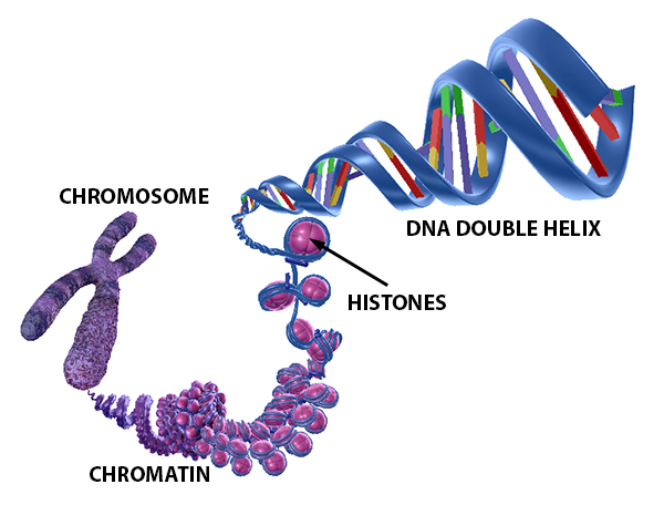 DNA structure and storage. Image from Visible Biology.
There are 46 separate strings of DNA in each somatic cell of the human body. Each one of these is called a chromosome. Scientists group them into 23 homologous pairs, which means that the chromosomes in each pair are similar in structure and function. The only exception to this is the 23rd pair—the sex chromosomes—in biologically male individuals. X and Y sex chromosomes only have certain regions (autosomal regions) that are homologous.
At the molecular level, DNA has a characteristic double-helix shape, and though this wasn’t observed by scientists until mid-20th century, it has quickly become one of the most iconic shapes in all of science.
Lesson on DNA structure from the Visible Biology YouTube series with Dr. Cindy Harley.
The sides of this twisted ladder are composed of alternating molecules of sugar (deoxyribose, to be precise) and a phosphate group. Each side is named for the direction it runs in (5’–3’ or 3’–5’). The ladder’s “steps” are composed of two nitrogenous bases, held together with hydrogen bonds.
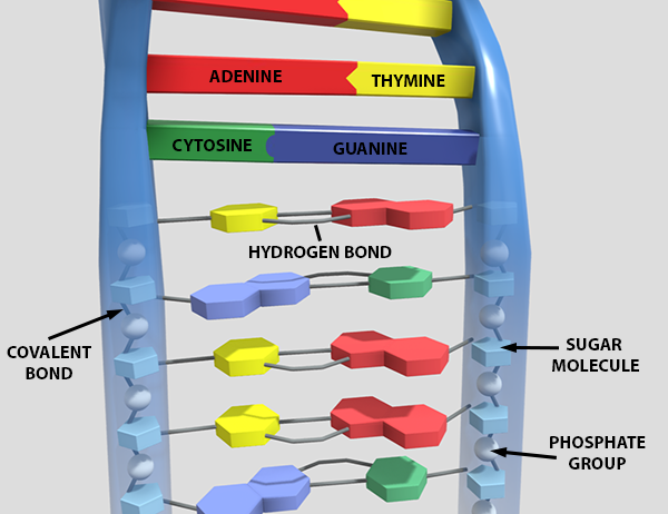 Molecular structure of DNA. Image from Visible Biology.
Four nitrogenous bases—cytosine, thymine, adenine, and guanine—can be found on strands of DNA. In terms of their chemical structure, cytosine and thymine are pyrimidines and adenine and guanine are purines. Adenine and thymine (A and T) always pair together, and guanine and cytosine (G and C) always pair together. They pair this way because A and T form two hydrogen bonds with each other and G and C form three.
At the most basic level, different sections of DNA strands (sequences of nitrogenous bases) provide instructions for the synthesis of proteins. A single section of DNA can even code for multiple proteins!
Replication: Doubling Up on DNA
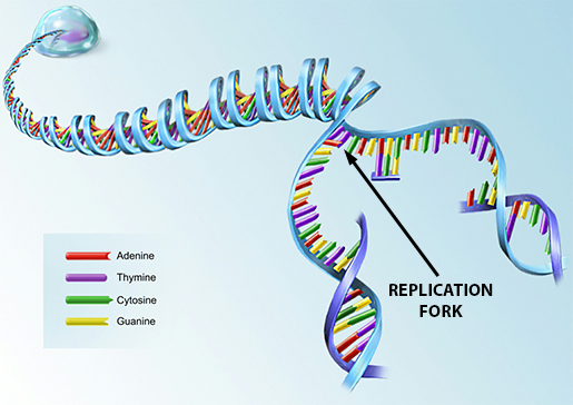 Illustration from A&P 6.
Replication of a cell’s DNA occurs before a cell prepares to undergo division—either mitosis or meiosis I.
It takes place in three(ish) steps.
- DNA unwinds from the histones.
- An enzyme called DNA helicase opens up the helix structure on a segment of DNA, breaking the bonds between the nitrogenous bases. It does this in a zipper-like fashion, leaving a replication fork behind it.
- Here’s where things get funky.
- On the 5’–3’ strand of the DNA, an enzyme called DNA polymerase slides towards the replication fork and uses the sequence of nitrogenous bases on that strand to make a new strand of DNA complementary to it (this means that its bases pair with the ones on the old strand).
- On the 3’–5’ strand, multiple DNA polymerases match up base pairs in partial segments, moving away from the replication fork. Later, DNA ligase connects these partial strands into a new continuous segment of DNA.
Want to know something neat? When a DNA molecule replicates, each of the resulting new DNA molecules contains a strand of the original, so neither is completely “new." Also, new histones are made at the same time the DNA replicates so that the new strands of DNA can coil around them.
Interlude: RNA vs DNA
Before we discuss transcription and translation, the two processes key to protein synthesis, we need to talk about another kind of molecule: RNA.
RNA is a lot like DNA—it’s got a sugar-phosphate backbone and contains sequences of nitrogenous bases. However, there are a couple of vital differences between RNA and DNA:
- RNA has only one nucleotide chain. It looks like only one side of the DNA ladder.
- RNA has ribose as the sugar in its backbone.
- RNA has Uracil (U) instead of thymine.
- RNA is smaller than DNA. RNA caps out at around 10,000 bases long, while DNA averages about 100 million.
- RNA can leave the nucleus. In fact, it does most of its work in the cytoplasm.
There are several different types of RNA, each with different functions, but for the purposes of this article, we’re going to focus on messenger RNA (mRNA) and transfer RNA (tRNA).
Making a Protein, Part 1: Transcription
Transcription is the first phase of the protein-making process, even though the actual protein synthesis doesn’t happen until the second phase. Essentially, what happens during transcription is that an mRNA “copies down” the instructions for making a protein from DNA.
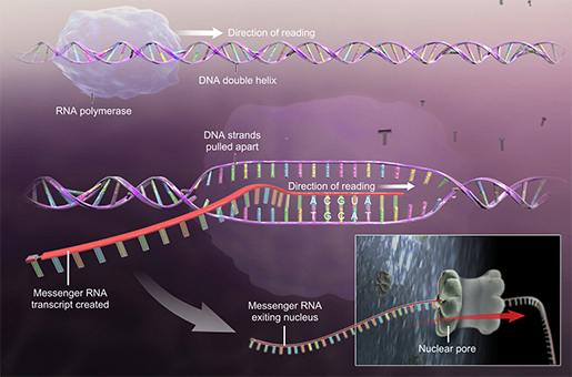 Illustration from A&P 6.
First, an enzyme called RNA polymerase opens up a section of DNA and assembles a strand of mRNA by “reading” the sequence of bases on one of the strands of DNA. If there’s a C on the DNA, there will be a G on the RNA (and vice versa). If there’s a T on the DNA, there will be an A on the RNA, but if there’s an A on the DNA, there will be a U (instead of a T) on the RNA. As the RNA polymerase travels down the string of DNA, it closes the helical structure back up after it.
Before the new mRNA can go out to deliver its protein fabrication instructions, it gets “cleaned up” by enzymes. They remove segments called introns and then splice the remaining segments, called exons, together. Exons are the sequences that actually code for proteins, so they’re the ones the mRNA needs to keep. You can think of introns like padding between the exons.
Also, remember how I mentioned that a single sequence of DNA can code for multiple proteins? Alternative splicing is the reason why: before the mRNA leaves the nucleus, its exons can be spliced together in different ways.
Lesson on transcription from the Visible Biology YouTube series with Dr. Cindy Harley.
Making a Protein, Part 2: Translation
After it’s all cleaned up and ready to go, the mRNA leaves the nucleus and goes out to fulfill its destiny: taking part in translation, the second half of protein construction.
Lesson on translation from the Visible Biology YouTube series with Dr. Cindy Harley.
In the cytoplasm, the mRNA must interface with tRNA with the help of a ribosome. tRNA is a type of RNA that has a place to bind to free amino acids and a special sequence of three nitrogenous bases (an anticodon) that binds to the ribosome.
Ribosomes are organelles that facilitate the meeting of tRNA and mRNA. During translation, ribosomes and tRNA follow the instructions on the mRNA and assemble amino acids into proteins.
Each ribosome is made up of two subunits (large and small). These come together at the start of translation. Ribosomal subunits can usually be found floating around in the cytoplasm, but a ribosome will dock on the rough endoplasmic reticulum if the protein it’s making needs to be put into a transport vesicle. Ribosomes also have three binding sites where tRNA can dock: the A site (aminoacyl, first position), the P site (peptidyl, second position) and the E site (the exit position).
Ultimately, translation has three steps: initiation, elongation, and termination.
During initiation, the strand of mRNA forms a loop, and a small ribosomal subunit (the bottom of the ribosome) hooks onto it and finds a sequence of bases that signals it to begin transcription. This is called the start codon (AUG).
Then, a tRNA with UAC anticodon pairs with this start codon and takes up the second position (P) site of the ribosome. This tRNA carries the amino acid Methionine (Met). At this point, the large ribosomal subunit gets in position as well (it’s above the mRNA and the small subunit is below).
In the elongation phase, the fully-assembled ribosome starts to slide along the mRNA. Let’s say the next sequence of bases it encounters after the start codon is GCU. A tRNA molecule with the anticodon CGA will bind to the first position (A) site of the ribosome. The amino acid it’s carrying (alanine) forms a peptide bond with Met. Afterward, the CGA tRNA (carrying the Met-Ala chain) moves to the second position and the UAC tRNA enters the E binding site. The first position site is then ready to accept a new tRNA. This process keeps going until the ribosome gets to a “stop” codon.
 Video footage from animation in Visible Biology.
Termination is pretty much what it sounds like. Upon reaching the “stop” codon, the tRNA that binds to the first position carries a protein called a release factor. The amino acid chain then breaks off from the ribosome, either going off into the cytosol or into the cisterna of the rough ER, and the ribosome disassembles. However, it might very well reassemble and go around the mRNA loop again. Also, multiple ribosomes can work on the same mRNA at once!
And those are the basics of DNA!
Here’s a handy chart you can look at if you need to remember the differences between transcription, translation, and replication:
|
Location |
Purpose |
Main Participants |
Product(s) |
|
|
Replication |
Nucleus |
Duplicate a full strand of DNA |
DNA |
2 identical strands of DNA |
|
Transcription |
Nucleus |
Use a strand of DNA to build a molecule of mRNA |
DNA |
mRNA |
|
Translation |
Cytoplasm |
Use mRNA to build an amino acid chain |
mRNA tRNA (and amino acids) |
Amino acid chain (protein) |
If you want to learn more about cells, check out these related VB Blog posts:
- Anatomy & Physiology: Parts of a Human Cell
- Tiny Transportation: Passive vs. Active Transport in Cells
For more information about the structure of DNA and chromosomes, check out our DNA eBook!
Be sure to subscribe to the Visible Body Blog for more anatomy awesomeness!
Are you an instructor? We have award-winning 3D products and resources for your anatomy and physiology course! Learn more here.
Additional Sources:
- Crash Course Biology #10: DNA Structure and Replication
- Crash Course Biology #11: DNA, Hot Pockets, & The Longest Word Ever
- Saladin (2015). Anatomy & Physiology: The Unity of Form and Function. 7th ed.
